
1.1 POJA-L604. Neutrophilic granulocyte
|
1.1 POJA-L604
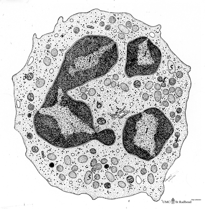
Title: Neutrophilic granulocyte Description: Scheme electron microscopy. The neutrophil is a phagocytic cell (12-15 µm) with a segmented lobular nucleus (3-5 lobes) and many cytoplasmic granules filled with degradative enzymes. These PMN cells (polymorph nuclear leukocytes) are the major cell types mediating acute inflammatory response to bacterial infections. The cytoplasm contains several types of granules (schematically only the two important types are depicted). (1): Nuclear lobes; (2): Golgi area; (3): Mitochondria; (4): Secondary or specific granules; (5): Lysosomes or primary granules. The neutrophilic granules are much smaller than the granules in eosinophils and basophils. |
Background: The neutrophil contains the following types of structurally and functionally different granules:
- The primary or azurophilic granules which are positive for myeloperoxidase. These granules are synthesized at the promyelocytic stage only. These granules do have little capacity for exocytosis. The granules contain: CD63, CD68, presenilin; elastase, cathepsin G, proteinase 3, defensins, BPI, myeloperoxidase (MPO), lysozyme; sialidase, azurocidin, β-glucuronidase.
- The secondary or specific granules which are negative for myeloperoxidase. The proteins in these granules are sythesized at the myelocyte stage only. These granules do have little capacity for exocytosis. These granules contain: CD11b/CD18, CD66, CD67, Gp91phox/p22phox; TNFRc, uPAR; SNAP-23, VAMP-2, stomatin, PGLYRP; collagenase, gelatinase, uPA, cystatin C and F; hCAP18, NGAL, B12BP, lysozym, lactoferrin, haptoglobin, pentraxin 3, prodefensin, α-1-antitrypsin, SLPI, orosomucoid, heparanase, β2-microglobulin, CRISP3.
- Granules with a high content of gelatinase are formed at the metamyelocyte stage and band cell stage, after which granule formation ceases. These granules contain CD11b/CED18, CD67, Gp91phox/p22phox,MMP25, TNFR; SNAP-23, VAMP-2, Nramp1; gelatinase, arginase-1; lysozyme; β2-microglobulin, CRISP3
- Secretory vesicles are then formed by endocytosis. They contain CD11b/CD18, CD66, CD67, Gp91phox/p22phox; MMP25; LIR1-4, -6, -7, -9; CD35; IFN-αR1 and IFN-αR2; TNFR1 and TNFR2; IL2;CXCR-4;CCR-1, -2; Ig (G,A,E)FcR; TLR-1,-MD2c; fMLPR; TREM1;SNAP-23, VAMP-2; plasmaproteins.
For a complete review the reader is referred to the paper “Neutrophil granules: a library of innate immunity proteins. Niels Borregaard, Ole E. Sørensen and Kim Theilgaard-Mönch. Trends in Immunology 28, (8), 340-345, 2007 “
The neutrophils circulate in the blood for only 6 hours and undergo programmed cell death if the cell is not recruited for an inflammation site. Neutrophils are recruited from the blood to sites of infection by binding to adhesion molecules (VLA’s, VCAM, LFA’s) on endothelial cells and by chemoattractants. They bind to microbes via receptors for mannose and opsonins (Ig, complement factors), Toll-like receptors (TLR’s). The ingested microbes are killed in phagolysosomes. The cells in fact generate a respiratory burst and produce reactive oxygen intermediates as well as reactive nitrogen intermediates as antimicrobial mediators. Cells have also high levels of defensins. The cells also express receptors for Ig, complement, Toll-receptors, mannose etc. to facilitate the phagocytosis.
Keywords/Mesh: blood, bone marrow, neutrophilic granulocyte, lysosome, azurophilic granule, primary granule, secondary granule, phagocytosis, histology, electron microscopy, POJA collection