
1.1 POJA-L853. Plasma cell with Russell bodies (nose septum, rat)
1.1 POJA-L853
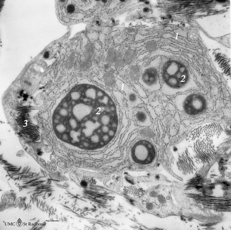
Title: Plasma cell with Russell bodies (nose septum, rat)
Description: Electron microscopy.
This mature plasma cell exhibits in the cytoplasm dilated rough endoplasmic reticulum (1) filled with electron-grey material as well as electron-dense granules (2) of varying sizes (antibodies). These electron-dense granules are made up of accumulated (crystallin) immunoglobulins within the lumen of the rough endoplasmic reticulum cisternae (so-called Russell bodies). Hyperstimulation can result in the formation of these Russell bodies, easily detectable in light microscopy.
(3): Collagen fibers in the nose septum.
Background: If the number of Russell bodies increases, the result will be a Mott cell found in pathologic states. Nucleus and Golgi areas are compressed by the enormous sacs containing Russell bodies. The cytoplasm almost disappears (e.g. in malignant tumours, parasitosis, hypergammaglobulinemias).
Keywords/Mesh: blood, bone marrow, plasma cell, Russell body, Mott cell, histology, electron microscopy, POJA collection