
4.1.1 POJA-L-3946+3945+3944+3942+3941. Immunohistochemical staining of the Plexus of Auerbach in duodenum (guinea pig)
4.1.1 POJA-L3946+3945+3944+3942+3941
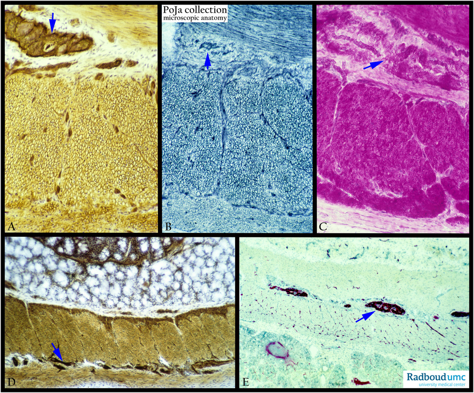
Title: Immunohistochemical staining of the Plexus of Auerbach in duodenum (guinea pig)
Description: Stain: (A) ATPase. (B) SDH. (C) Phosphorylase. (D) ATPase. (E) AFFA.
The Plexus of Auerbach or myenteric plexus (arrows, ↘↘) is located between the inner circular muscle layer and the outer longitudinal muscle layer in the intestinal tract. The plexus consists of ganglion cells surrounded with mantle layer cells (glia cells) and nerve strands that innervate both muscle layers and thus regulate the contraction and movement of the intestines. Note that also the layers muscle fibers are stained positively (A, B, C, and D).
In (E), however the ganglion cells are much more conspicuous than the muscle fibers.
Keywords/Mesh: small intestine, Plexus of Auerbach, ganglion cells, immunohistochemistry, ATPase, phosphorylase, histology, POJA collection.