
5.4.1 POJA-L5005+La0045+
5008+5009+2313+2527.
Glomerulus (I) in the kidney
5.4.1 POJA-L5005+La0045+5008+5009+2313+2527
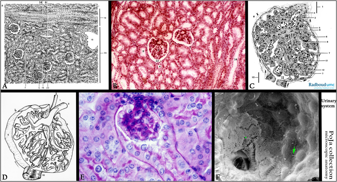
Title: Glomerulus (I) in the kidney
Description:
(A): Kidney cortex, scheme, human.
(5) Glomerulus. (16) Afferent arteriole.
(6) Arteria corticalis radiata (cortical radial artery). (18) Vena arcuata (arcuate vein).
(7) Urinary pole where the urinary space empties into the first part (19) Medulla radiata (medullar rays).
of the tubular system, pars contorta I (proximal tubule).
(9) Pars contorta I (proximal tubule). (20) Cortical labyrinth.
(14) Collecting tubule. (22) Bowman’s space.
(B): Kidney cortex, stain modified Azan, human.
(4) Pars contorta I (distal tubule). Other numbers are equal to (A).
(C, D): Glomerulus, schemes, human.
(1) Vascular pole. (7) Podocyte (visceral epithelial cell).
(2) Vas afferens or afferent arteriole. (8) Endothelial cell.
(3) Vas efferens. (9) Mesangium cell.
(4) Goormaghtigh cells/extraglomerular mesangium cells (lacis cells). (10) Urinary pole, (proximal convoluted tubule, pars contorta I).
(5) Bowman’s capsule. (11) Bowman’s space.
(6) Epithelial (parietal) cell of the glomerular capsule. (12) Capillary.
(E): Glomerulus, PAS stain, mouse. Glomerulus and urinary pole showing that the PAS-positive material is localized along the basement membranes of tubules and blood vessels in- and outside the glomerulus. The glomerular mesangium is deeply stained.
Note, however, that also the microvilli of the proximal tubules are strongly positive since they are coated with a glycoprotein layer.
(F): Scanning electron microscopy of the urine pole of the glomerulus, perfusion-fixed, rat. Note that the cells of the proximal convoluted tubule (PCT) are studded with microvilli (*). The one-layered flattened parietal cells (Bowman’s capsule) with the bulging nuclei and one single cilium (arrow) per cell are well presented.
Keywords/Mesh: urinary system, kidney, glomerulus, vascular pole, urinary pole, Bowman’s capsule, PAS, histology, electron microscopy, POJA collection