7.1 POJA-L1210+1332
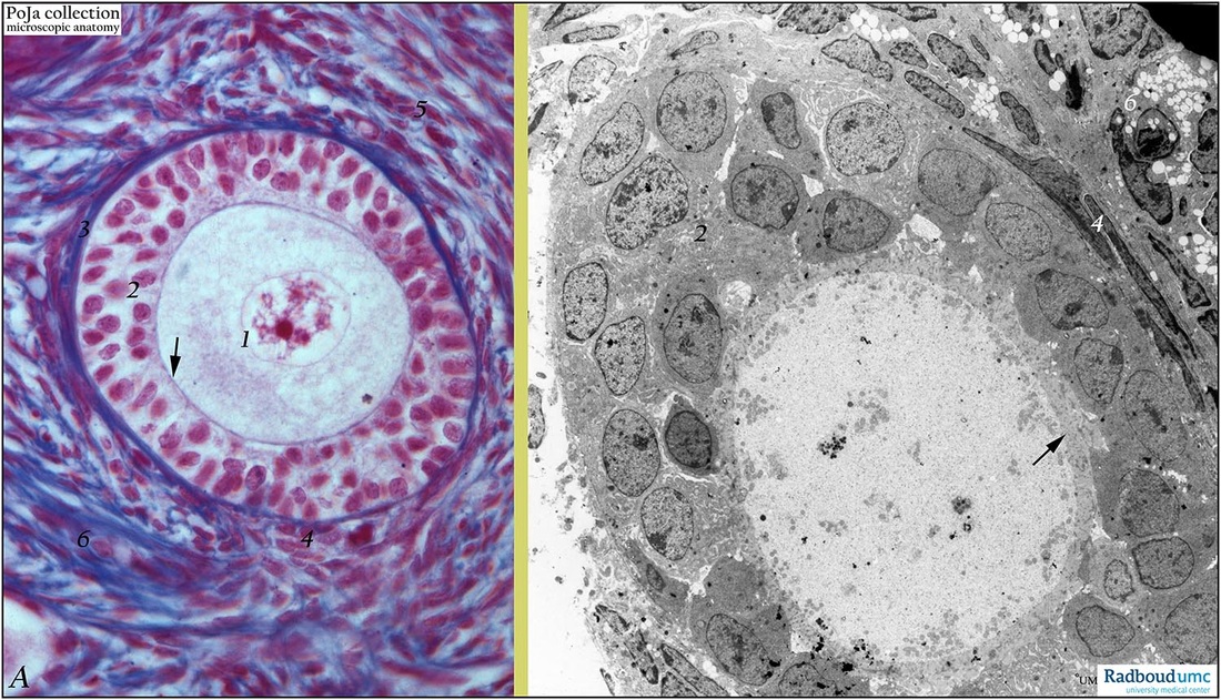
Title: Secondary follicle in ovary (rabbit)
Description: (A) Stain: Azan. (B) Electron microscopy (gerbil).
(A): Type 3 secondary follicle (pre-antral follicle) surrounded by 2-3 layers of granulosa (follicular) cells. Note vesicular aspect of nucleus (1) with distinct nucleolus in oocyte. A thin (bluished) zona pellucida (↑) is visible in close contact with cuboidal granulosa cells (2). Granulosa cells are devoid of blood capillaries and therefore are rich in cellular contact and communication. The border between follicle and ovarian stroma is formed by a membrana vitrea, a thick blue-stained basement membrane reinforced with collagen and reticular fibers (3). Close to the latter differentiating stroma cells form at a few places the internal theca (4) consisting of spindle-like cells and blood capillaries. At (5) the external theca (i.e. theca externa) as the most outer layer starts to develop. The stroma cells and collagen fibers (6) encompass the follicle.
(B): Ultra structurally a similar follicle with (arrow) zone pellucida surrounding large electron-light oocyte cytoplasm (nucleus is not sectioned). Two-three layers of granulosa cells (2) and intercellular dilated spaces filled with liquor folliculi. Note close association of inner ring of granulosa cells with a relatively thick basal lamina-like zona pellucida. Elongated internal theca cells (4), and vacuolated cells are interstitial cells (6).
Keyword/Mesh: female reproductive organs, ovary, ovarian follicle, granulosa cells, theca cells, female genitalia, theca cells, granulosa cells, histology, POJA collection

