
4.2.1 POJA-L3788+3787+3789+3790. Examples of cholestasis in the liver (human)
4.2.1 POJA-L-3788+3787+3789+3790
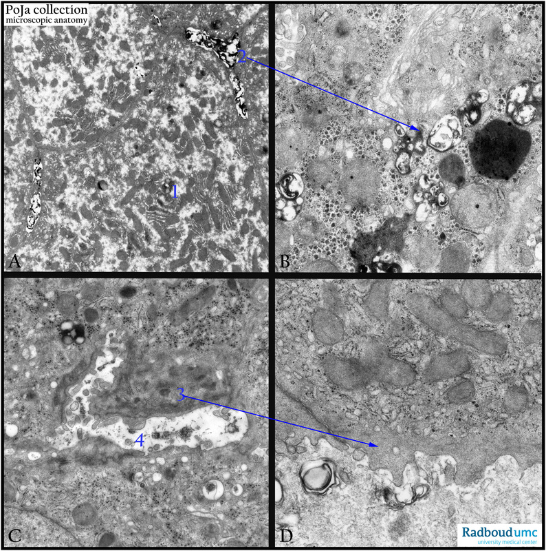
Title: Examples of cholestasis in the liver (human)
Description: Electron micrographs.
(A, 2) Canalicular bile plugs between individual hepatocytes or within bile ducts may also be seen, representing bile that has been excreted from the hepatocytes but cannot go any further due to the obstruction.
The accumulation of bile products (A, 1 and detail in B, 2) is seen as liposome-like structures.
(C, D, 3) The wall of an obstructed bile canaliculus (4) or ductules can be “reinforced” or thickened by aggregates of filaments. The pile up of bile can occur in the canaliculi but also within the liver cell leading eventually to apoptosis or necrosis.
Keywords/Mesh: liver, cholestasis, bile plugs, electron microscopy, POJA collection