12.1.3 POJA-L2534+2552+2957+2551
Title: Iris and ciliary body
Description:
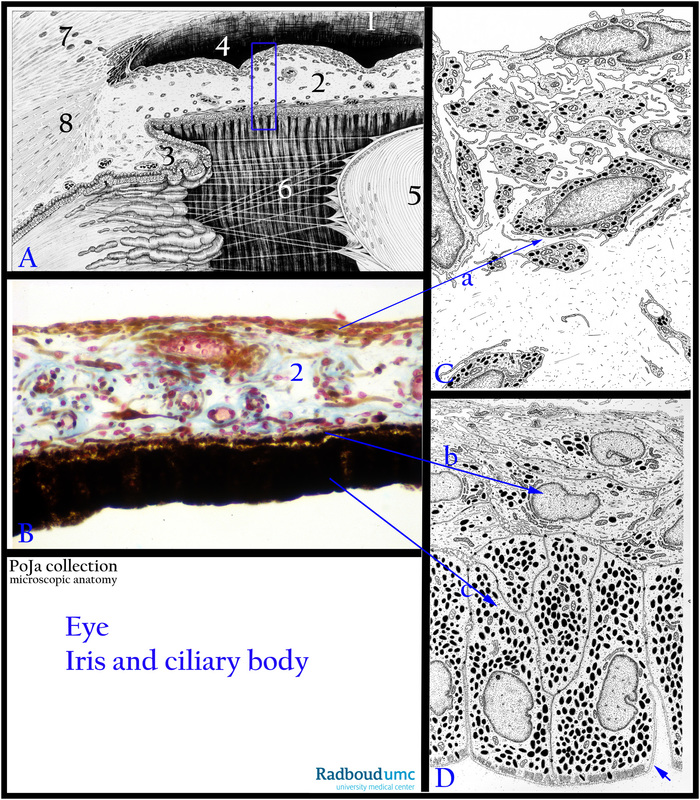
(A, C, D): Electron microscopy schemes, human.
(A): (1) Cornea.
(2) Iris.
(3) Ciliary body and ciliary processes.
(4) Anterior chamber.
(5) Lens, covered with a single epithelial layer and a capsule of collagen IV and glycosaminoglycans.
(6) Suspensory ligament or zonular fibres arise from the ciliary body.
(7) Limbus corneae (or limbal area) with Schlemm canal.
(8) Ciliary muscle close to the sclera.
(B): The iris (blue rectangle A,2) is detailed here, stain trichrome Mallory, monkey. The iris is well vascularised and also contains
pigmented melanocytes and pigmented phagocytes.
The front side of the iris is detailed in (C)(arrow 2a towards (C). This front side of the iris is directed towards the anterior chamber,
is not covered with an epithelial lining but the surface is formed by mutually connected fibroblasts and some pigmented cells.
The posterior side of the iris is detailed in (D) with arrows (2b) and (2c). This posterior side consists of a double layer of epithelial cells,
i.e. the outer layer contains numerous pigment granules (melanin). It is bordered by a basal lamina (arrow) towards the vitreous body.
The inner layer with a basal lamina borders the connective stroma and contains much less melanin. These cells possess contractile extensions running parallel to the surface in a radial orientation (myoepithelial cells). The cells are interconnected with each other
as well as with the pigmented epithelial cells by junctional complexes. Their contractile extensions form the dilator papillae muscle
(arrow 2b from (B) to (D).
(A second muscle, called sphincter papillae muscle comprises concentric arranged smooth muscle tissue in the stroma near the pupillary border (not shown here).
Keywords/Mesh: eye, iris, ciliary body, lens, histology, electron microscopy, POJA collection

