5.4.1 POJA-L2364+2369+2539+2362+2360+2541
Title: Podocyte in glomerulus (X) of the kidney
Description:
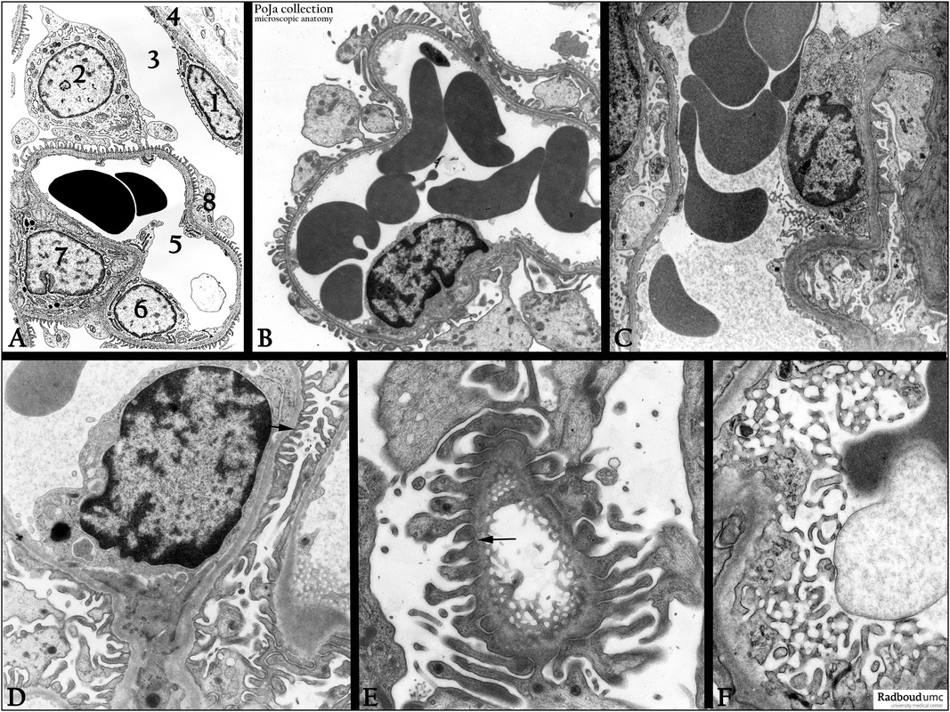
(A): Glomerulus, electron microscopy scheme, human.
- Epithelial cell (parietal cell) (1) lining the Bowman’s capsule (4).
- Podocyte (2) that engulfs the blood capillary with small finger-like primary and secondary processes (8) that surround the cpillary (5).
- Primary urine space (Bowman space) (3).
- Endothelial cell (6) lining the lumen (5).
- Mesangium cell (7).
(B-F): Electron microscopy of equivalent structures as shown in (A), rat. Note the pedicles and the filtration slit membranes
between them in (D, E) (arrows). (F) Illustrates the tangential sectioned fenestrated endothelial cells in the glomerular capillaries.
Keywords/Mesh: urinary system, kidney, glomerulus, capillary, podocyte, filtration slit membrane, fenestrated endothelium, histology, electron microscopy, POJA collection

