
4.2.1 POJA-L3729+3677. Sinusoid endothelial cell and hepatic stellate cell in liver sinusoid (rat
4.2.1 POJA-L3729+3677
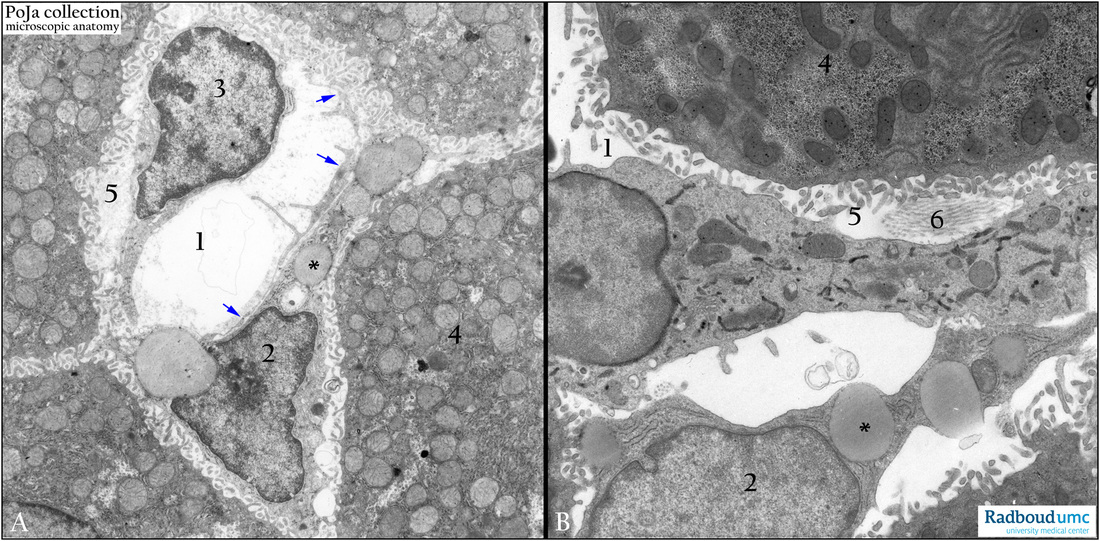
Title: Sinusoid endothelial cell and hepatic stellate cell in liver sinusoid (rat)
Description: Electron micrograph of survey (A) and detail (B) of cells in liver sinusoid (peroxidase staining with DAB).
(1) Liver sinusoid. In (B) a Kupffer cell partly covers the space of Disse. This phagocyte is characterized by the presence of peroxidase in its RER. Hepatic stellate cell (fat-storing cell), (2) is localized between the lining sinusoid endothelial cell (3) and the liver cell (4), i.e. within the space of Disse (5). These hepatic stellate cells (cells of Ito) contain fat droplets (*). Note that in the space of Disse fibers (6) of the reticular meshwork are found. The arrows (↘) point to the extreme slender processes of the covering sinusoid endothelial cell. They cover therefore the fat storing cell.
Keywords/Mesh: liver cell, hepatic stellate cell, sinusoid endothelial cell, Kupffer cell, space of Disse, electron microscopy, POJA collection