
13.1 POJA-L4711+La-0314+4675
Lymphatic capillaries (human) in lymph node and digestive tract
13.1 POJA-L4711+La-0314+4675
Title: Lymphatic capillaries (human)
Description:
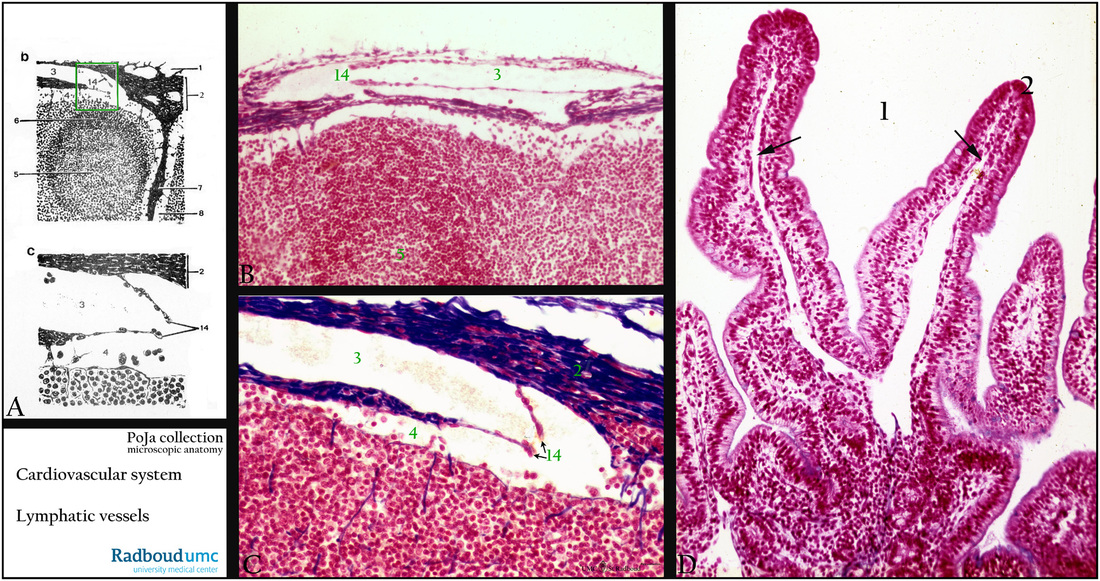
(A, b+c): Scheme lymph node. (1) Fat cells. (2) Capsule. (3) Afferent lymphatic vessel into the lymph node. (5) Active germinal centre with lymphocytes and antigen-presenting cells. (6) Lunula of the follicle. (14) Flap-like valves in the afferent lymphatic vessel. (7) Trabecula of connective tissue. (8) Intermediate penetrating sinus.
(B, C): Azan stain, lymph node and lymphatic vessel. Identical structures as shown in (A).
(D): Azan stain, lymphatic capillary (arrows) in villi of small intestine. These single blind-ending capillaries are also known as lacteals. (1) Lumen, (2) villus.
Background: The lymphatic capillary network acts as a drainage system removing fluid or lymph from tissue. The lymphatic capillaries and vessels pass also through the lymph nodes and convey lymphocytes (and antigens) to be processed and activated in the lymph follicle. Blind-ending lymphatic capillaries are responsible for the absorption of the interstitial tissue fluid. When interstitial tissue fluid enters the initial lymphatic capillaries the fluid is called lymph and contains i.a. surplus of interstitial tissue fluid, waste products, circulating white blood cells. Lymph vessel structure is similar to blood capillaries and can display also fenestrae or gaps. There are more than 500 lymph nodes in the human body, located along the lymphatic system at variable intervals. Afferent lymph vessels carry the clear lymph into the marginal sinus of a lymph node, where it percolates through the reticular network of the node and then it is drained by one efferent lymph vessel.
Keywords/Mesh: cardiovascular system, vascularisation, lymphatic system, lymph node, germinal center, lymph, lymphatic capillary,, small intestine, valve, lacteal, histology, POJA collection
Title: Lymphatic capillaries (human)
Description:
(A, b+c): Scheme lymph node. (1) Fat cells. (2) Capsule. (3) Afferent lymphatic vessel into the lymph node. (5) Active germinal centre with lymphocytes and antigen-presenting cells. (6) Lunula of the follicle. (14) Flap-like valves in the afferent lymphatic vessel. (7) Trabecula of connective tissue. (8) Intermediate penetrating sinus.
(B, C): Azan stain, lymph node and lymphatic vessel. Identical structures as shown in (A).
(D): Azan stain, lymphatic capillary (arrows) in villi of small intestine. These single blind-ending capillaries are also known as lacteals. (1) Lumen, (2) villus.
Background: The lymphatic capillary network acts as a drainage system removing fluid or lymph from tissue. The lymphatic capillaries and vessels pass also through the lymph nodes and convey lymphocytes (and antigens) to be processed and activated in the lymph follicle. Blind-ending lymphatic capillaries are responsible for the absorption of the interstitial tissue fluid. When interstitial tissue fluid enters the initial lymphatic capillaries the fluid is called lymph and contains i.a. surplus of interstitial tissue fluid, waste products, circulating white blood cells. Lymph vessel structure is similar to blood capillaries and can display also fenestrae or gaps. There are more than 500 lymph nodes in the human body, located along the lymphatic system at variable intervals. Afferent lymph vessels carry the clear lymph into the marginal sinus of a lymph node, where it percolates through the reticular network of the node and then it is drained by one efferent lymph vessel.
Keywords/Mesh: cardiovascular system, vascularisation, lymphatic system, lymph node, germinal center, lymph, lymphatic capillary,, small intestine, valve, lacteal, histology, POJA collection