5.7 POJA-La0092+La0080+L2438+4255+2439+2495+2440
Title: Facet cells in the urothelium
Description:
Electron microscopy.
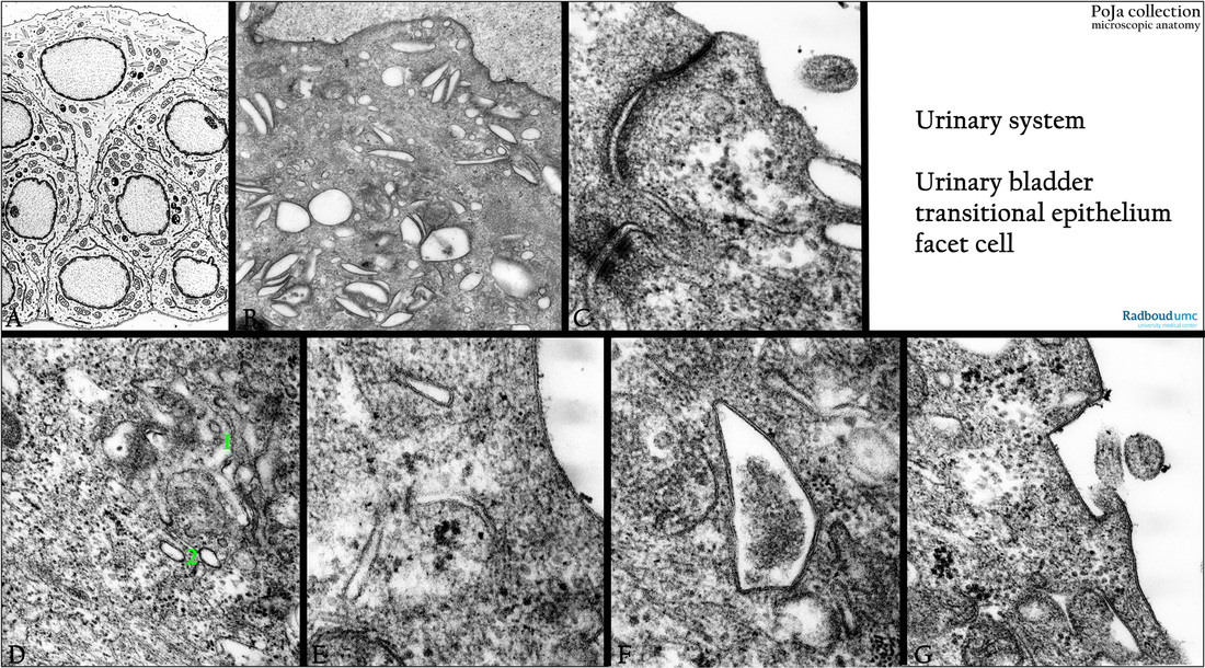
(A): Scheme of superficial cells or facet cells in urothelium, human.
(B): Facet cell with larger round as well as smaller fusiform or discoid vesicles containing the plaques. gerbil.
The rigid-looking plaques are also known as the asymmetrically thickened membrane domains (or AUMs).
In between apical bundles of sectioned intermediate-sized filaments.
(C - G): Facet cells, human.
Junctional complexes between facet cells (C).
Golgi area (1) in facet cell with small fusiform vesicles (2), that are produced in the post-Golgi compartments (D).
Facet cell with apical fusiform vesicles with AUMs (E).
Facet cell with large AUM vesicle enclosing granulofilamentous debris (F).
Surface urothelial cells with incorporation of AUM at the cell surface (hinge area) (G).
The plaques occurring in the mentioned discoid vesicle membranes contribute to continuous apical membrane regeneration because the vesicles act as transporting compartments to deliver urothelial plaques to the apical cell membrane.
These specialized vesicles (each containing two plaques) can be added and removed from the cell membrane quickly as the bladder expands or contracts.
The apical plasma membrane of the facet cells is covered with mannosylated uroplakin protein particles.
Uroplakins are transmembrane proteins acting as a special barrier to a.o.water, small non-electrolytes.
The AUM consists of the assembly of the four uroplakins Ia, Ib, II and III. Together with the tight junctions of the junctional complexes these plaques create a most impermeable barrier for the urine.
Background: It is shown that plaques form gradually in the individual post-Golgi compartments as uroplakin-positive transporting vesicles, immature fusiform vesicles and mature fusiform vesicles. The plaques are AUMs with diameters between 500- 1500 nm separated by thin rims of non-thickened membrane called the hinge regions.
Members of the Rab family like 11a , 27b as well as syntaxin, SNAP-23 and synaptobrevin play an important role in the apical targeting
of fusiform/discoid vesicles. During stretching of the bladder mechanoreceptors activate exocytosis of the fusiform/discoid vesicles
and are modulated by a.o. cAMP, Ca2+, extracellular ATP, epidermal growth factor receptors etc..
Keywords/Mesh: urinary bladder, urothelium, transitional epithelium, facet cell, fusiform vesicle, discoid vesicle, AUM, uroplakin, histology, electron microscopy, POJA collection

