
14.1 POJA-L6032B+6102 Mitochondria localisation in myofibres
14.1 POJA-L6032B+6102 Mitochondria localisation in myofibres
14.1 POJA-L6032B+6102 Mitochondria localisation in myofibres
Title: Mitochondria localisation in myofibres
Description:
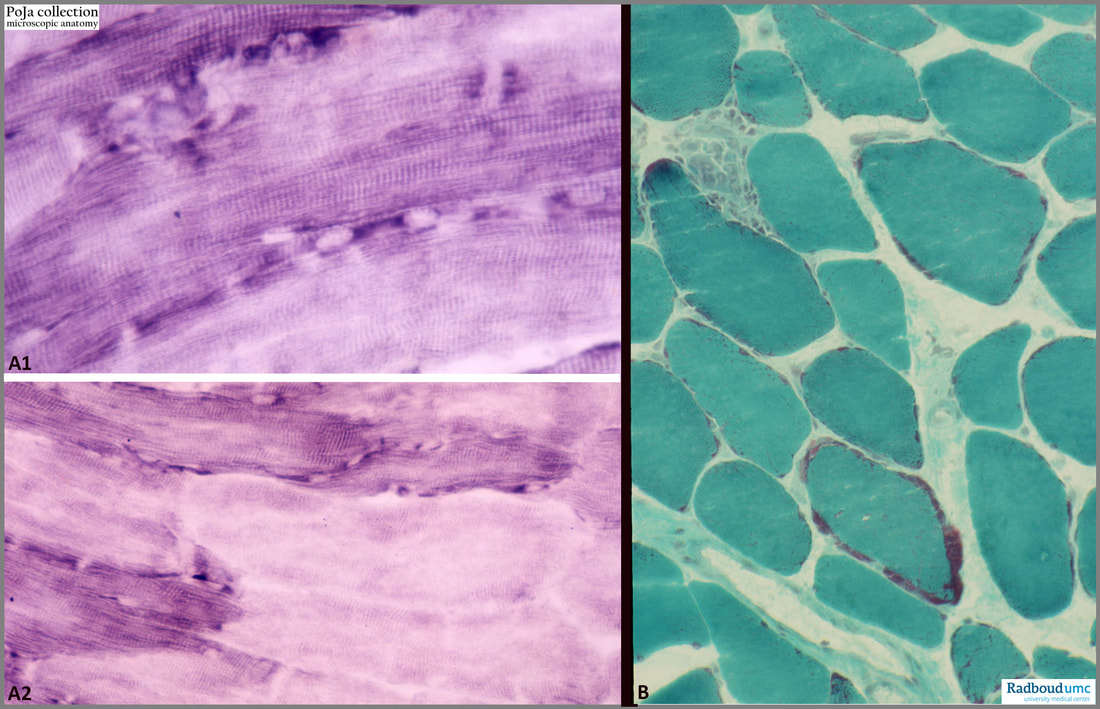
(A1-A2): NADH-TR staining of mitochondria in longitudinal sectioned skeletal muscles. Type I (red) fibres are positively stained, type II (white) fibres are stained less or negative (rat). Around the unstained nuclei the deep purple-blue stain indicates many mitochondria. The striations also stain positive due to the localisation of mitochondria within the sarcomeres. Type I fibres (rich in mitochondria) are slow-twitch oxidative motor units, while type II fibres generally are fast-twitch oxidative glycolytic motor units.
(B): Trichrome staining showing numerous fine dots over the cross-sectioned muscle fibres, and peripheral purple-red spots of accumulated mitochondria (mouse).
See also:
Keywords/Mesh: locomotor system, skeletal muscle, striated muscle, type I fibre, type II fibre, oxidative enzyme, mitochondrion, histology, POJA collection
Description:
(A1-A2): NADH-TR staining of mitochondria in longitudinal sectioned skeletal muscles. Type I (red) fibres are positively stained, type II (white) fibres are stained less or negative (rat). Around the unstained nuclei the deep purple-blue stain indicates many mitochondria. The striations also stain positive due to the localisation of mitochondria within the sarcomeres. Type I fibres (rich in mitochondria) are slow-twitch oxidative motor units, while type II fibres generally are fast-twitch oxidative glycolytic motor units.
(B): Trichrome staining showing numerous fine dots over the cross-sectioned muscle fibres, and peripheral purple-red spots of accumulated mitochondria (mouse).
See also:
Keywords/Mesh: locomotor system, skeletal muscle, striated muscle, type I fibre, type II fibre, oxidative enzyme, mitochondrion, histology, POJA collection