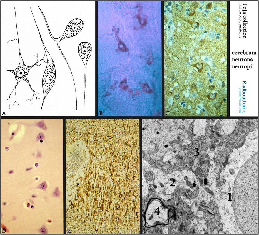
11.5 POJA-L3014+3020+3021+3038+3044+3056
Title: Nerve cells and glial cells in cerebrum
Description:
(A): Scheme of several types of nerve cells, i.e. multipolar-, bipolar, pseudo unipolar cells and unipolar cells, human.
(B): Acetylcholine-esterase staining of pyramidal cells in cat.
(C): Immunoperoxidase staining of neuron specific-enolase (NSE) with DAB, human. Neurons react positively
while astroglial cells remain negative.
(D): Stain hematoxylin-eosin, human. Neurons in cortex, some of them contain lipofuscin. Astroglial cells are shown as smaller 'naked' nuclei.
(E): Immunoperoxidase staining with DAB and antibodies against neurofilament (NF) in medulla, human. At the left an arteriole surrounded by a wide paravascular space (Virchow-Robin) as a para-arterial influx route for cerebrospinal fluid to enter the brain parenchyma. (See: Glymphatic System in POJA-L4750)
(F): Electron micrograph of part of a neuron embedded in neuropil, human. (1) Neuron with nucleus. (2) Dendrite.
(3) Neuropil. Note the electron-dense stained myelin of axon (4).
(C/ E, by courtesy of H. ter Laak PhD, Department of Pathology, University Medical Centre Radboud University, Nijmegen,
The Netherlands).
Keywords/Mesh: nervous tissue, cerebrum, acetylcholine-esterase, neuron specific-enolase, neuropil, neuron, axon, dendrite, neurofilament, histology, electron microscopy, POJA collection

