
4.1.1 POJA-L0345+3547+3551. Pancreatic acinus with intercalated duct (human, rabbit)
4.4.1 POJA-L0345+3547+3551
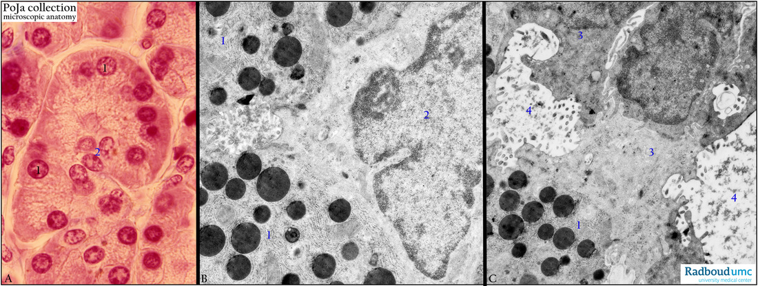
Title: Pancreatic acinus with intercalated duct (human, rabbit)
Description: Stain (A) Azan. (B, C) Electron micrograph.
The acini of the exocrine pancreas consist of secretory epithelial cells (1) arranged centrally around an intercalated duct (A, 2, B 2 and C, 3) starting with a centroacinar cell (B 2, C 3) that exports the secreted zymogen granules eventually to an interlobular duct and subsequently to the duodenum. (C, 4) represents the lumen (with fluffy secretion products) between the secretory cells and a centroacinar cell at the beginning of an intercalated duct. Note that zymogen granules are electron-dense and are located between the rough endoplasmic reticulum.
Keyword/Mesh: pancreas, exocrine, acinus, intercalated duct, electron microscopy, histology, POJA collection