
2.2 POJA-L975. Scheme of blood circulation in the red pulp in the spleen
2.2 POJA-L975
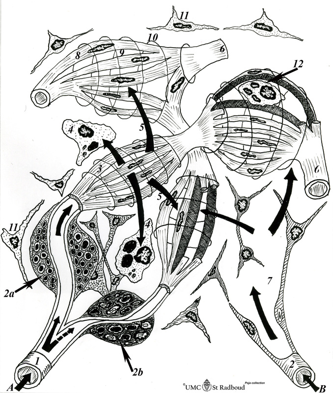
Title: Scheme of blood circulation in the red pulp in the spleen
Description:
(A): The route of "closed circulation" proposes that blood empties from the capillaries directly into the splenic sinus. The central arteriole bifurcates into penicillar arterioles (1) and the blood slowly enters ensheathed capillaries, surrounded by aggregations of macrophages (2a/b) and a network of reticular cells and fibers, and drains into the splenic cords and into sinusoids (3). The blood leaves the sinusoids through split spaces (3) between the endothelial cells and leak into the venous sinuses, Aged red cells with rigid cell membranes are lysed and destroyed and removed by phagocytes (4) along the sinus walls. Malaria-infected red blood cells are pitted by these macrophages. Healthy erythrocytes recirculate back into the sinuses (5) and leave the system via a pulpa vein (6) and trabecular veins (e.g. in dogs and rats)
(B): The route of "open circulation" proposes that the blood empties into spaces (7) between reticular cells of the red pulp outside the sinus and then enters the circulation through slits in the sinus wall (5, 3). About 90% of the blood takes the open route of circulation. Blood enters a macrophage-ensheathed capillary (2) and reaches more efficiently the marginal sinuses in the marginal zone where blood cells come into contact with dendritic antigen-presenting cells (APC) resulting into trapping of foreign antigens to be presented to appropriate lymphocytes. Subsequently blood drains back via slit spaces between the endothelial cells into the sinusoids and leaves the system via a pulpa vein (6) as it is mostly the case in human.
(C): Details of a spleen sinus
8. Endothelial cells of a sinus
9. Slit-like spaces between the lining cells,
10. Sinus wall enclosed by reticular fibers (argyrophilia)
11. Reticular cells
12. Phagocytising endothelial cell
Keywords/Mesh: lymphatic tissue, spleen, red pulp, open circulation, closed circulation, histology, POJA collection