14.1 POJA-L6162 Electron micrograph of a cross sectioned sarcomere of skeletal muscle fibre (human)
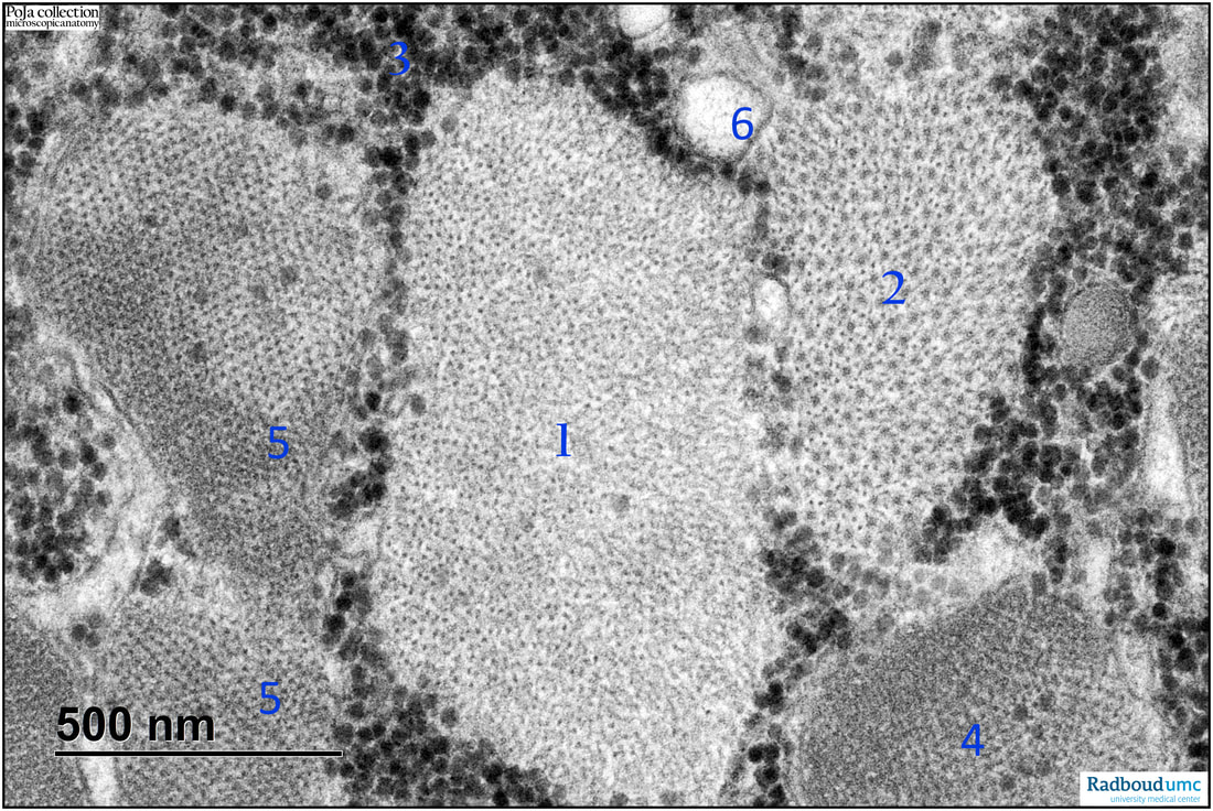
14.1 POJA-L6162 Electron micrograph of a cross sectioned sarcomere of skeletal muscle fibre (human)
(Micrographs by courtesy of L. Eshuis BSc Section Electron microscopy, Department of Pathology, Radboud university medical center, Nijmegen The Netherlands)
Title: Electron micrograph of a cross sectioned sarcomere of skeletal muscle fibre (human)
Description: The electron micrograph shows an arbitrary cross section through the sarcomere.
- (1): At I-band level.
- (2): At A-band level.
-(3): Glycogen deposits (including dispersed granules within the various band regions).
-(4): Z-line region.
-(5): M-region.
-(6): Sarcoplasmic reticulum.
See also:
Keywords/Mesh: locomotor system, skeletal muscle, striated muscle, sarcomere, A-band, I-band, Z-line, sarcoplasmatic reticulum, glycogen, electron microscopy, POJA collection
Title: Electron micrograph of a cross sectioned sarcomere of skeletal muscle fibre (human)
Description: The electron micrograph shows an arbitrary cross section through the sarcomere.
- (1): At I-band level.
- (2): At A-band level.
-(3): Glycogen deposits (including dispersed granules within the various band regions).
-(4): Z-line region.
-(5): M-region.
-(6): Sarcoplasmic reticulum.
See also:
- 14.1 POJA-L6223A Cross section human muscle myofibrils
Keywords/Mesh: locomotor system, skeletal muscle, striated muscle, sarcomere, A-band, I-band, Z-line, sarcoplasmatic reticulum, glycogen, electron microscopy, POJA collection

