
5.4.3 POJA-L2449+2432+
2455+2456+2457.
Transition from connecting tubule (CNT) into cortical collecting duct (CCD) (XV) of the human kidney
5.4.3 POJA-L2449+2432+2455+2456+2457
Title: Transition from connecting tubule (CNT) into cortical collecting duct (CCD) (XV) of the human kidney
Description:
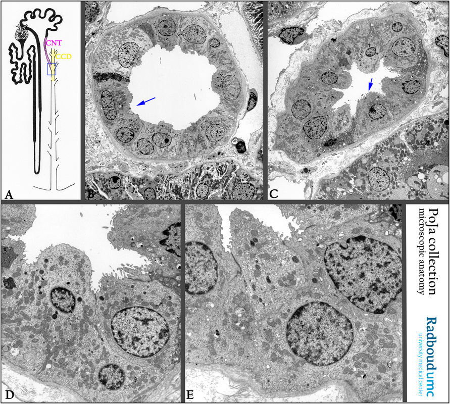
(A): Scheme nephron. The blue rectangle focusses to the transitional zone from the connecting tubule (CNT) to the cortical collecting
duct (CCD).
Electron micrographs of CNTs.
(B): CCD. Arrow points to two intercalated cells with numerous apical vesicles.
(C): Transition from CNT to CCD, close to a PCT in the cortex. Arrow points to an intercalated cell.
(D): The CNT cells differ from the CCD cells. They possess a basal labyrinth and more mitochondria throughout the cytoplasm.
However intercalated cells normally are also present in both CNT and CCD segments.
(E): The transition segment contains both CNT proper cells and intercalated cells.
Keywords/Mesh: urinary system, kidney, renal tubule, connecting tubule, CNT, collecting duct, CCD, histology, electron microscopy, POJA collection
Title: Transition from connecting tubule (CNT) into cortical collecting duct (CCD) (XV) of the human kidney
Description:
(A): Scheme nephron. The blue rectangle focusses to the transitional zone from the connecting tubule (CNT) to the cortical collecting
duct (CCD).
Electron micrographs of CNTs.
(B): CCD. Arrow points to two intercalated cells with numerous apical vesicles.
(C): Transition from CNT to CCD, close to a PCT in the cortex. Arrow points to an intercalated cell.
(D): The CNT cells differ from the CCD cells. They possess a basal labyrinth and more mitochondria throughout the cytoplasm.
However intercalated cells normally are also present in both CNT and CCD segments.
(E): The transition segment contains both CNT proper cells and intercalated cells.
Keywords/Mesh: urinary system, kidney, renal tubule, connecting tubule, CNT, collecting duct, CCD, histology, electron microscopy, POJA collection