
9.5 POJA-L3520+3531+3533
+3534+3508+3512.
Nerve network (IV) in the medulla of the rat adrenal gland
9.5 POJA-L3520+3531+3533+3534+3508+3512
Title: Nerve network (IV) in the medulla of the rat adrenal gland
Description:
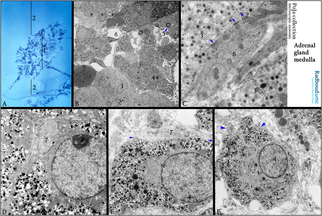
Adrenal gland. (A): The acetylcholine esterase activity as a marker for nerve endings indicate the presence of an ample nerve fiber
network in the medulla (1), while the cortical area (2) remains devoid.
Electron microscopy of the adrenal gland (B-F).
(B): Shows both adrenalin (3) and noradrenalin (4) containing cells in the medullary zone. (5) Indicates the myelinated axons.
(6) is a sinusoid with an endothelial cell. It is thought that noradrenalin cells seem to be contacted by denser AChE-reactive nerve
fibers than adrenalin cells.
(C): Reveals the granules of adrenalin cells (3) that show close synaptic contacts (arrows) with cholinergic axons (6).
(D): Represents noradrenalin cells with numerous small electron-dense granules, and a close synaptic contact (7) with a preganglionic sympathetic axon.
(E): A noradrenalin cell (4). Between the two arrows synapses (7) and several preganglionic axon endings.
(F): A noradrenalin cell (4) and axon endings with synapses (arrows). (8) Resident macrophage with numerous lysosomes.
Keywords/Mesh: adrenal gland, medulla, adrenalin, noradrenalin, nerve cell, synaps, cholinergic axon, histology, electron microscopy, POJA collection
Title: Nerve network (IV) in the medulla of the rat adrenal gland
Description:
Adrenal gland. (A): The acetylcholine esterase activity as a marker for nerve endings indicate the presence of an ample nerve fiber
network in the medulla (1), while the cortical area (2) remains devoid.
Electron microscopy of the adrenal gland (B-F).
(B): Shows both adrenalin (3) and noradrenalin (4) containing cells in the medullary zone. (5) Indicates the myelinated axons.
(6) is a sinusoid with an endothelial cell. It is thought that noradrenalin cells seem to be contacted by denser AChE-reactive nerve
fibers than adrenalin cells.
(C): Reveals the granules of adrenalin cells (3) that show close synaptic contacts (arrows) with cholinergic axons (6).
(D): Represents noradrenalin cells with numerous small electron-dense granules, and a close synaptic contact (7) with a preganglionic sympathetic axon.
(E): A noradrenalin cell (4). Between the two arrows synapses (7) and several preganglionic axon endings.
(F): A noradrenalin cell (4) and axon endings with synapses (arrows). (8) Resident macrophage with numerous lysosomes.
Keywords/Mesh: adrenal gland, medulla, adrenalin, noradrenalin, nerve cell, synaps, cholinergic axon, histology, electron microscopy, POJA collection