11.2.2 POJA-L2076+3280+3281+4310+4311+3282+3283
Title: Sensory receptors (Vater-Pacini/Meissner's corpuscles)
Description:
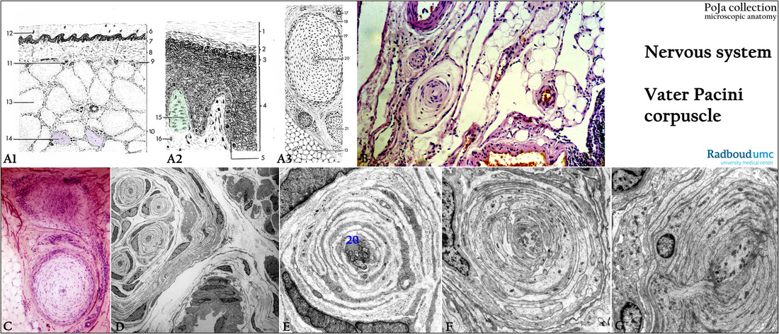
(A1, A2, A3): Scheme of localization and structure of the sensory receptors (afferent nerve ending) in the skin of finger (tactile corpuscle
of Meissner) and foot sole (lamellated corpuscle of Vater-Pacini, Pacinian corpuscle), human.
(A1): Skin of the soles of the feet. (A2): Skin of the finger. (A3): Pacinian corpuscle. (1) Stratum corneum. (2) Stratum lucidum. (3) Stratum granulosum. (4) Stratum spinosum. (5) Stratum basale. (6) Epidermis. (7) Stratum papillare. (8) Stratum reticulare. (9) Stratum glandulo-vesiculare. (10) Subcutis. (11) Sweat glands in stratum glandulo-vasculare. (12) Duct of a sweat gland. (13) Adipose tissue. (14) Pacinian corpuscle. (15) Meissner’s corpuscle. (16) Myelinated nerve; (17) Small artery. (18) Sheath of connective tissue. (19) Connective tissue lamellae of the perineural capsule. (20) Thickened central nerve fiber. (21) Nerve bundle. See also Section 10
(B): Stain hematoxylin-eosin, human. The Pacinian corpuscle is located in the subcutis of the skin.
(C): Stain Weigert-hematoxylin, human. Pacinian corpuscles located in the adipose tissue of the skin
(D, E): Electron micrographs of Pacinian corpuscles, gastrocnemius biopsy, human.
(F): Electron micrograph of Pacinian corpuscle, snout of pig
(G): Electron micrograph of Pacinian corpuscle, snout of cat.
Background: The Pacinian corpuscles are up to 4 mm long and 2 mm thick and in most mammals they are identical. They are located in the subcutaneous adipose tissue of the soles of the feet, the palms of the hand but also near the fasciae, periosteum and tendon, in the vicinity of arteriovenous anastomoses as well as in the pancreas and mesenteric places. The sheaths consist of concentric layers of cytoplasmic processes (perineural lamellae) of Schwann cells. Between the lamellae the spaces are liquid-filled, and also contain few capillaries and thin collagen fibrils. The sensory nerve axon is located centrally and terminates in a nodular thickening (20) containing many mitochondria. The whole is surrounded with a capsule of elastic fiber network continuous with the perineurium. The corpuscles (mechanoreceptors) are sensitive for vibration and changes in pressure.
Keywords/Mesh: nervous tissue, sensory receptor, sensory nerve ending, Vater Pacini corpuscle, pacinian corpuscle, Meissner’s corpuscle, skin, histology, electron microscopy, POJA collection

