15.1 POJA-L7008+7020 Foetal hyaline cartilage 1
15.1 POJA-L7008+7020 Foetal hyaline cartilage 1
Title: Foetal hyaline cartilage 1
Description:
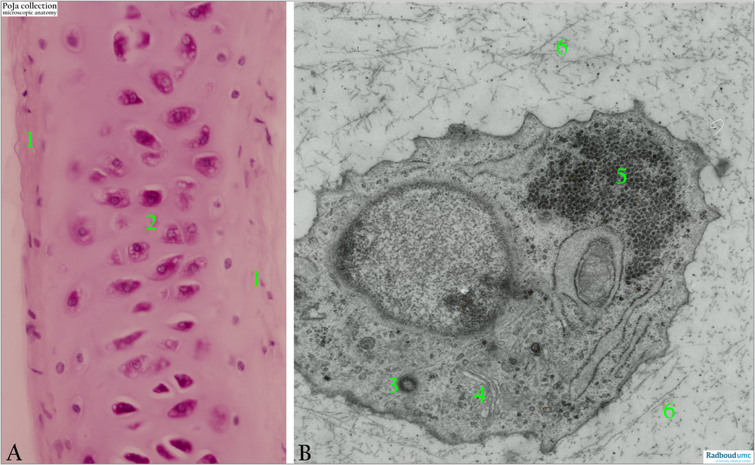
Foetal human hyaline cartilage, Haematoxylin-eosin staining (A) and electron micrograph (B).
(1): Perichondrium.
(2): Lacunar chondrocytes.
(3): Centriole.
(4): Golgi area.
(5): Glycogen.
(6): Matrix. (collagen fibres and dense proteoglycan particles).
Background:
Hyaline cartilage develops from mesenchymal blastema. Mesenchyme cells differentiate into chondroblasts that form cartilage (matrix, proteoglycans, glycoproteins, and collagen II fibrils becoming masked by the matrix material). They further differentiate into chondrocytes that lay in the lacunae or cartilage caverns. By interstitial growth the chondrocytes divide within the lacunae and are designated as isogenous islets of cells. In the shown example the isogenous cell groups are not yet formed.
The cartilage matrix is rich in e.g. collagen, proteoglycans (PG) and chondronectin.
Generally the matrix is composed of 10-30% collagen, 3%-10% PG and other minor glycoproteins and lipids.
PG molecules are composed of protein cores covalent linked to heparan sulphate, chondroitin 6-sulphate/chondroitin 4-sulphate and are highly anionic charged. The negative charges attract cations and especially sodium ions which in turn attract water molecules. The matrix is hydrated in that extent (60-85% of the wet weigh is water) that cartilage is able to resist compression. Furthermore the side chains of the PG bind electrostatically to collagen resulting in a cross-linked framework of collagen fibres and ground substance that can resist tensile forces.
The large molecule chondronectin has binding sites for collagen type II, hyaluronic acid, chondroitin 4-/chondroitin 6-sulphates and integrins, and in this way cells are attached to the fibrous and amorphous components of the matrix.
Reference:
Gartner Cartilage and Bone in Textbook of Histology 2021 ISBN:9780323672726
See also:
- 15.0 POJAL7009+7021 Foetal hyaline cartilage 2
Keywords/Mesh: locomotor system, cartilage, hyaline, foetus, matrix, chondrocyte, collagen, proteoglycan, electron microscopy, histology, POJA collection

