4.2.1 POJA-L-3738+3742+3743
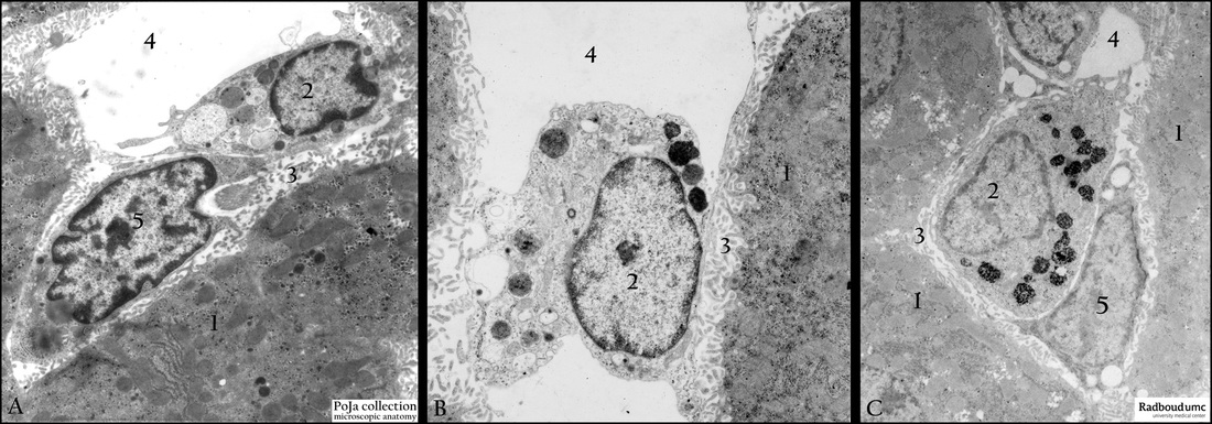
Title: Kupffer cells and hepatic stellate cells in liver (rat)
Description: Electron micrographs. (A) Kupffer cell. (2) Lining the microvilli of the liver cells (1) and the space of Disse (3). The Kupffer cell is well filled with phagocytized material and lysosomes. (5) Hepatic stellate cell or cell of Ito localized in the space of Disse.
(B) The Kupffer cell has turned into a swollen cell still slightly attached with the processes to the environment. Note the conspicuous lysosomes.
(C) After ferritin perfusion and reinforced zinc oxide treatment the dark granular particles can be retrieved in the lysosomes of the Kupffer cells due to their strong phagocytosis capacity. (4) Sinusoid space. (5) Hepatic stellate cell or cell of Ito (fat-storing cell).
Keywords/Mesh: liver cell, Kupffer cells, hepatic stellate cells, ferritin, lysosomes, phagocytosis, electron microscopy, POJA collection

