2.1 POJA-L929-930
Title: The effect of cyclophosphamide treatment on the resident macrophages in thymus (rat)
Description: Stain: Immunoperoxidase DAB.
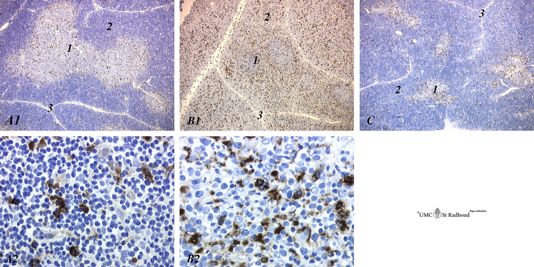
A single injection with cyclophosphamide (CP, 70 mg/ml) induces a transient cortical involution after 4 days, i.e. the darkly stained cortex and the lightly stained medulla in normal thymus (A1, A2) turn to darkly stained medulla and a lightly stained cortex (B1, B2) four days after a single injection with CP. The thymus recovers to normal about 10 days later (C).
The monoclonal antibody ED-1 stains the rat homologue of human CD68, a single chain glycoprotein of 110kDa that is expressed predominantly on the lysosomal membrane of tissue macrophages (and myeloid cells).
(A1, A2): Normal thymus showing most resident macrophages localised in the medulla (A2) (A1-1; A2), dark brown cytoplasmic stained macrophages in a lightly stained medulla). The darkly stained cortex (A1-2) contains much less resident macrophages.
(B1, B2): Four days after CP treatment the macrophages have occupied equally the depleted cortex and the darker stained medulla.
(C): Ten days later the status has been almost recovered to normal distribution of macrophages and lymphocytes.
(3): Shows septa or trabeculae dividing the thymus in lobes.
Keywords/Mesh: lymphatic organs, thymus, ED1 macrophages, cyclophosphamide, rat, toxicology, immunosuppression histology, POJA collection

