
11.4 POJA-L3095+3098+
3113+2970+3119+2969+4448+4449.
Oligodendroglial cells and astrocytes in cerebellum
11.4 POJA-L3095+3098+3113+2970+3119+2969+4448+4449
Title: Oligodendroglial cells and astrocytes in cerebellum
Description:
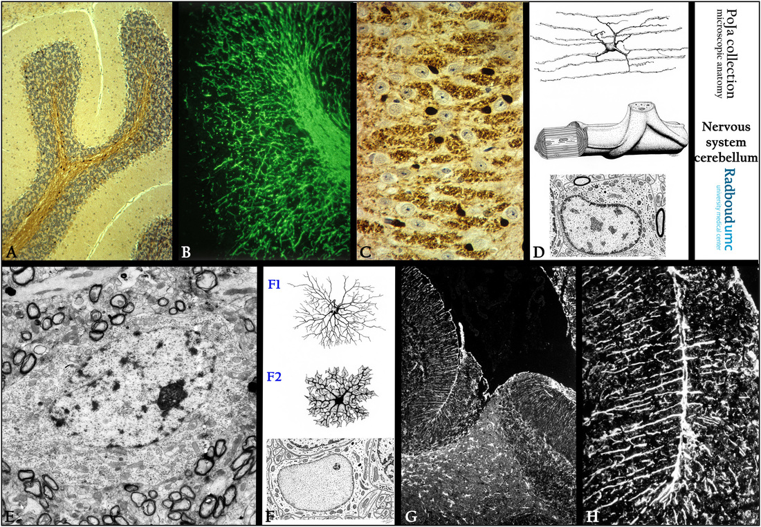
(A): Immunoperoxidase staining of the cerebellum with DAB and antibodies against carboanhydrase, mouse.
This enzyme is involved in the energy metabolism and show in survey strong presence in the scattered oligodendrocytes (dark brown-stained) of the medulla and granular layer.
(B): Imunofluorescence staining with antibodies against myelin basic protein (MBP) illustrating the presence of myelinated fibers in medulla and granular layer, mouse.
(C): Immunoperoxidase staining with DAB against carboanhydrase highlighting in the granular layer the close association
of the dark brown-stained oligodendrocytes and cross-sections of myelinated structures, mouse.
(A/B/C, by courtesy of H. ter Laak PhD, Department of Pathology, University Medical Centre of the Radboud University, Nijmegen,
The Netherlands).
(D): Electron microscopy scheme of oligodendroglia, human. Oligodendrocytes are characterized by having a fine network of parallel running delicate processes located between the rows of axon bundles. They form myelin sheaths around the nerve fibers by wrapping several times around the nerve fibers. One oligodendrocyte can myelinate up to 40 different axons and several internodes per axon.
The node is contacted by processes from perinodal astrocytes.
(E): Electron micrograph of a small neuron in the granular layer, monkey. The cell is surrounded by numerous myelinated and nonmyelinated nerve fibers.
(F): Electron microscopy scheme of astrocyte, human. The small bundles of intracellular glial filaments are intermediate-sized filaments that mostly react with antibodies against glial fibrillary acidic protein (GFAP).
The protoplasmic asterocytes (F2) are present in the gray matter Their terminal feet end upon capillaries at one side by formation of perivascular endfeet. At the other side they end up on neurons.
Additionally several astrocytes create, by subpial endfeet together with a basal lamina (membrana gliae limitans superficialis) a partition between the nervous system and the connective tissue of the pia mater.
The fibrous asterocyte (F1) generally resides in the white matter and just like the protoplasmic ones their processes also form several perivascular endfeet or subpial endfeet.
(G, H): Immunofluorescence staining with antibodies against the intermediate filament GFAP (glial fibrillary acidic protein), black- white, mouse. Positive reactivity is present in astroglial cells and its processes which terminate at the surface as glia limiting membrane (membrana gliae limitans superficialis).
Keywords/Mesh: nervous tissue, cerebellum, oligodendrocyte, myelination, myelin basic protein, carboanhydrase, histology, electron microscopy, POJA collection
Title: Oligodendroglial cells and astrocytes in cerebellum
Description:
(A): Immunoperoxidase staining of the cerebellum with DAB and antibodies against carboanhydrase, mouse.
This enzyme is involved in the energy metabolism and show in survey strong presence in the scattered oligodendrocytes (dark brown-stained) of the medulla and granular layer.
(B): Imunofluorescence staining with antibodies against myelin basic protein (MBP) illustrating the presence of myelinated fibers in medulla and granular layer, mouse.
(C): Immunoperoxidase staining with DAB against carboanhydrase highlighting in the granular layer the close association
of the dark brown-stained oligodendrocytes and cross-sections of myelinated structures, mouse.
(A/B/C, by courtesy of H. ter Laak PhD, Department of Pathology, University Medical Centre of the Radboud University, Nijmegen,
The Netherlands).
(D): Electron microscopy scheme of oligodendroglia, human. Oligodendrocytes are characterized by having a fine network of parallel running delicate processes located between the rows of axon bundles. They form myelin sheaths around the nerve fibers by wrapping several times around the nerve fibers. One oligodendrocyte can myelinate up to 40 different axons and several internodes per axon.
The node is contacted by processes from perinodal astrocytes.
(E): Electron micrograph of a small neuron in the granular layer, monkey. The cell is surrounded by numerous myelinated and nonmyelinated nerve fibers.
(F): Electron microscopy scheme of astrocyte, human. The small bundles of intracellular glial filaments are intermediate-sized filaments that mostly react with antibodies against glial fibrillary acidic protein (GFAP).
The protoplasmic asterocytes (F2) are present in the gray matter Their terminal feet end upon capillaries at one side by formation of perivascular endfeet. At the other side they end up on neurons.
Additionally several astrocytes create, by subpial endfeet together with a basal lamina (membrana gliae limitans superficialis) a partition between the nervous system and the connective tissue of the pia mater.
The fibrous asterocyte (F1) generally resides in the white matter and just like the protoplasmic ones their processes also form several perivascular endfeet or subpial endfeet.
(G, H): Immunofluorescence staining with antibodies against the intermediate filament GFAP (glial fibrillary acidic protein), black- white, mouse. Positive reactivity is present in astroglial cells and its processes which terminate at the surface as glia limiting membrane (membrana gliae limitans superficialis).
Keywords/Mesh: nervous tissue, cerebellum, oligodendrocyte, myelination, myelin basic protein, carboanhydrase, histology, electron microscopy, POJA collection