7.1 POJA-L1207+L1266+1903
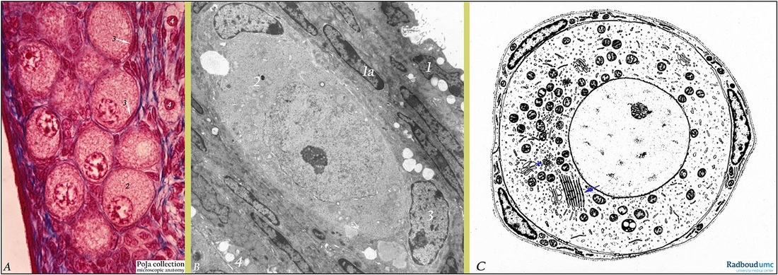
Title: Primordial follicles in ovary (rabbit, gerbil, human)
Description: (A) Stain: Azan (rabbit); (B) Electron microscopy (gerbil). See also scheme in POJA-L1201+l1231. (C) Scheme electron microscopy (human).
(A): Cortex contains many primordial follicles (type 1) just below flat surface epithelial lining (specialized peritoneal mesothelial cells, →1). Between follicles and epithelial lining exists a small rim of connective tissue of tunica albuginea. Oocytes (2) have large nuclei with distinct nucleoli. They are surrounded by a single layer of flattened follicular (or pregranulosa) cells (→ 3). Most of the stroma cells are spindle-like shaped with collagen fibers (blue) in between; two vacuolar cells (4) represent interstitial cells that produce small amounts of androgenic hormones.
(B): Flattened vacuolized mesothelial cells (1) separated by fibroblasts (1a) of tunica albuginea from a primordial follicle (type 1) with oocyte (2) Two follicular cells (3) and vacuolated interstitial cells are present (4).
(C): Scheme primordial follicle (type 1), compare with electron microscopy in B. Small mitochondria, small Golgi areas (*), annulate lamellae (→), lysosomes. Three flattened follicular (pregranulosa) cells encompass the oocyte; a basal lamina surrounds the follicle.
Background: Annulate lamellae are composed of parallel arrays of flattened cisternae and closely resemble the structure of nuclear envelopes. These membrane systems are often found as intracytoplasmic structures juxtanuclearly in embryonic cells a. o. oocytes, tumor cells. They are thought to play a role in cell growth and differentiation.
Keyword/Mesh: female reproductive organs, ovary, ovarian follicle, female genitalia, primordial follicle, rabbit, gerbil, human, interstitial cells, mesothelial cells,histology, POJA collection

