
13.1 POJA-L4592+4600+4632+4601+4635+4636+La0297 Arterioles
13.1 POJA-L4592+4600+4632+4601+4635+4636+La0297
Title: Arterioles
Description:
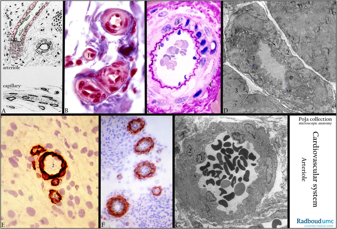
(A): Scheme arteriole and capillary. Capillary (6-30 micrometers): lumen with endothelium and pericytes. Arteriole (20-120 micrometers) longitudinal and cross-section: lumen with endothelium (green) and media (pink). The tunica media (= middle tunic) contains up to 1-2 concentric layers of smooth muscle cells (SMC).
(B): Arterioles and capillary (omentum), Azan, human. (1) Capillary. The other blood vessels are arterioles, note one or few SMCs in the media.
(C): Small-sized muscular artery (muscle biopsy), 1-micrometer plastic section PAS stain (perjood acid Schiff), human. (1) Endothelium with (2) strongly stained IEL (internal elastic lamella) due to strong reacting glycoproteins ensheathing the elastic lamina. (3) Media with SMC stains weakly due to minor presence of glycogen. (4) The adventitia stains strongly because the amount of ECM with all glycoproteins appears larger.
(D): Electron micrograph of arteriole, mouse. (1) Endothelial cell with numerous pinocytotic vesicles. Arrows point to the “junctional” contacts of endothelial basal processes piercing the basal lamina and touching the smooth muscle cells. (2) Smooth muscle cells. (3) Surrounding skeletal muscle tissue.
(E): Collagen IV in vascular basement membranes (nervous tissue, cerebrum), immunoperoxidase AEC- stained anti-collagen IV antibodies- Haematoxylin (frozen section), rat 1-day postnatally. Small capillary (1) and larger capillary (2) contain basement membrane collagen IV as part of the Blood-Brain barrier (BBB). Nuclei of neuroglia cells of the neuropil (nervous tissue without neuronal nuclei) are stained with Haematoxylin.
(F): alpha-Sm actin in blood vessels (middle ear region), immunoperoxidase AEC- stained anti-actin antibodies-Haematoxylin (frozen section), rat 4-day postnatally. Actin is detected in the contractile pericytes of capillaries as well as in smooth muscle cells in the media of larger vessels.
(G): Arteriole (biopsy nose region), EM, human. Lumen with erythrocytes. (1) Endothelium, (2) smooth muscle cells (SMC) with swollen mitochondria (due to artifactual procedures), (3) damaged adventitia.
Background: Arterioles (also called precapillary arteries) are the smallest arteries with a diameter of 20-120 micrometers. The intima comprises endothelial cells resting on a basement membrane (basal lamina) and an internal elastic lamina (IEL). The endothelium is continuous. The arteriolar media comprises one or two layers of smooth muscle cells (SMC). In the smallest arterioles the endothelial cells have basal processes which pierce the basal lamina and make junctional contacts with the SMCs. The adventitia consists of variable dense to loose connective tissue.
In the range from small arteries toward capillaries the terminal arteriole (or precapillary) loses its IEL and the media consists of one layer of circular arranged SMCs. The following segment is the metarteriole where SMCs disjoint and eventually replaced by pericytes. This segment is an important local regulator of blood flow and in this well-innervated area regulation of the peripheral resistance takes place.
Small arteries followed by arterioles, capillaries and draining venules together create the terminal flow circuit (‘Endstrombahn’). (See also: https://www.khanacademy.org/science/health-and-medicine/circulatory-system/blood-vessels/v/pre-capillary-sphincters)
Keywords/Mesh: cardiovascular system, vascularisation, blood vessel, capillary, pericyte, smooth muscle cell, actin, basal lamina, collagen IV, arteriole, metarteriole, muscular artery, histology, electron microscopy, POJA collection
Title: Arterioles
Description:
(A): Scheme arteriole and capillary. Capillary (6-30 micrometers): lumen with endothelium and pericytes. Arteriole (20-120 micrometers) longitudinal and cross-section: lumen with endothelium (green) and media (pink). The tunica media (= middle tunic) contains up to 1-2 concentric layers of smooth muscle cells (SMC).
(B): Arterioles and capillary (omentum), Azan, human. (1) Capillary. The other blood vessels are arterioles, note one or few SMCs in the media.
(C): Small-sized muscular artery (muscle biopsy), 1-micrometer plastic section PAS stain (perjood acid Schiff), human. (1) Endothelium with (2) strongly stained IEL (internal elastic lamella) due to strong reacting glycoproteins ensheathing the elastic lamina. (3) Media with SMC stains weakly due to minor presence of glycogen. (4) The adventitia stains strongly because the amount of ECM with all glycoproteins appears larger.
(D): Electron micrograph of arteriole, mouse. (1) Endothelial cell with numerous pinocytotic vesicles. Arrows point to the “junctional” contacts of endothelial basal processes piercing the basal lamina and touching the smooth muscle cells. (2) Smooth muscle cells. (3) Surrounding skeletal muscle tissue.
(E): Collagen IV in vascular basement membranes (nervous tissue, cerebrum), immunoperoxidase AEC- stained anti-collagen IV antibodies- Haematoxylin (frozen section), rat 1-day postnatally. Small capillary (1) and larger capillary (2) contain basement membrane collagen IV as part of the Blood-Brain barrier (BBB). Nuclei of neuroglia cells of the neuropil (nervous tissue without neuronal nuclei) are stained with Haematoxylin.
(F): alpha-Sm actin in blood vessels (middle ear region), immunoperoxidase AEC- stained anti-actin antibodies-Haematoxylin (frozen section), rat 4-day postnatally. Actin is detected in the contractile pericytes of capillaries as well as in smooth muscle cells in the media of larger vessels.
(G): Arteriole (biopsy nose region), EM, human. Lumen with erythrocytes. (1) Endothelium, (2) smooth muscle cells (SMC) with swollen mitochondria (due to artifactual procedures), (3) damaged adventitia.
Background: Arterioles (also called precapillary arteries) are the smallest arteries with a diameter of 20-120 micrometers. The intima comprises endothelial cells resting on a basement membrane (basal lamina) and an internal elastic lamina (IEL). The endothelium is continuous. The arteriolar media comprises one or two layers of smooth muscle cells (SMC). In the smallest arterioles the endothelial cells have basal processes which pierce the basal lamina and make junctional contacts with the SMCs. The adventitia consists of variable dense to loose connective tissue.
In the range from small arteries toward capillaries the terminal arteriole (or precapillary) loses its IEL and the media consists of one layer of circular arranged SMCs. The following segment is the metarteriole where SMCs disjoint and eventually replaced by pericytes. This segment is an important local regulator of blood flow and in this well-innervated area regulation of the peripheral resistance takes place.
Small arteries followed by arterioles, capillaries and draining venules together create the terminal flow circuit (‘Endstrombahn’). (See also: https://www.khanacademy.org/science/health-and-medicine/circulatory-system/blood-vessels/v/pre-capillary-sphincters)
Keywords/Mesh: cardiovascular system, vascularisation, blood vessel, capillary, pericyte, smooth muscle cell, actin, basal lamina, collagen IV, arteriole, metarteriole, muscular artery, histology, electron microscopy, POJA collection