7.1 POJA-L1767C+1768C+1946+1949
Title: Brenner tumor, ovary (human, adult)
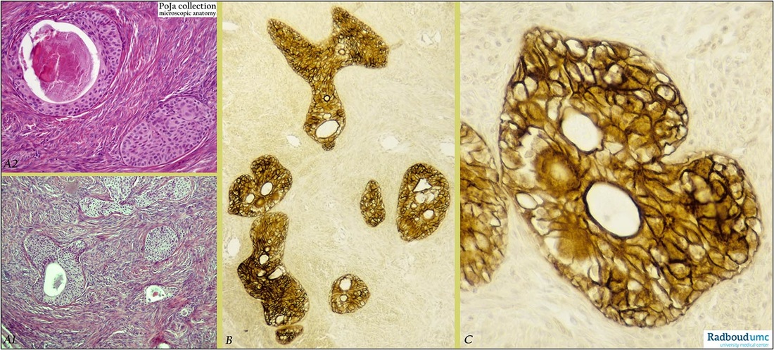
Description: Stain: (A) Hematoxylin-eosin; (B, C) CK7 (OVTL12/30) antikeratin antibody immunoperoxidase staining with diaminobenzidin reaction (DAB) and hematoxylin counterstaining.
(A1): Low magnification of fibroepithelial tumor composed of ovarian-derived stroma. Nests of transitional cells present with focal humanization and containing focally mucin.
(A2): Higher magnification of similar tumor with well demarcated epithelial nests in fibrostromal part. The nest is composed of transitional-like cells (bladder urothelium-like) with a central cyst containing debris and mucin.
(B): Several CK7 positive well-demarcated epithelial nests and some branching columns. Small cystic spaces are also present. Fibrous stroma is CK7-negative.
(C): Higher magnification reveals compact epithelial arrangement of CK7-positive cell nest. In this picture cystic spaces are lined by cuboidal cells. Stroma is CK7-negative. (Figure A1, A2: By courtesy of F. van de Molengraft MD PhD, Department of Pathology, Rijnstate Hospital Arnhem, The Netherlands)
Background: In most cases Brenner tumors are benign but borderline (proliferative Brenner tumor) and malignant types are also reported. The Brenner tumor belongs to the surface epithelial-stromal tumors and is a transitional cell tumor of the ovary. It is assumed that the Brenner tumor develops from the ovarian serosa but that this type of tumor is formed of urothelium and was differentiated along the Wolffian pathway. It is known that in the embryo the coelomic epithelium overlies the nephrogenital ridge and cells from that epithelial area over this ridge retain the capacity to differentiate along that line. Alternatively it was recently suggested that the origin from transitional epithelial nests might be located at paraovarian sites (Kurman and Shih 2010). Brenner tumors appear as solid as well as cystic. The epithelial component of Brenner tumors consists of sharply demarcated nests of transitional cells resembling those lining the urinary bladder. These cell nests might contain microcysts or glandular spaces lined by centrally columnar or mucin-secreting cells. Generally normal urothelium of the human urinary bladder expresses heterogeneous CK7 reactivity. However in painful bladder syndrome and in a variety of urothelial cancers the urothelium shows in most cases an increase in expression of CK7. In case of Brenner tumors expression of an overall uniform strong CK7 (OVTL12/30 and RCK105) reactivity in paraffin-embedded sections is noted. This result is in line with the observations that during abnormal differentiation all transitional tumor cells express CK7.
Keywords/Mesh: female reproductive organs, ovary, Brenner tumor, female genitalia,, ovarian neoplasms, keratin 7, histology, pathology
POJA collection

