7.1 POJA- L1215+L1313+L1317
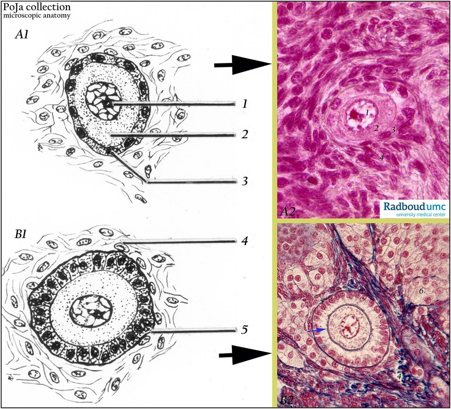
Title: Primordial and primary follicles in ovary (scheme, rabbit)
Description: (A1, B1) Scheme (human). Stain: (A2) Azocarmine G; (B2) Azan.
(A1): Scheme (human): primordial follicle (type 1). Nucleolus (1). Ooplasm (2). Flattened layer of follicular (pregranulosa) cells (3).
(A2): Rabbit: intermediary primordial follicle (late type 1) with vesicular nucleus and distinct nucleolus (1) in a granular ooplasm (2). A single layer of mixed elongated and cuboidal follicle cells surrounds oocyte (3). Cell-rich ovarian stroma of cortex (4).
(B1): Scheme (human): unilaminar primary follicle (type 2). Cell-rich ovarian stroma of cortex (4). Basement membrane (membrana vitrea) (5).
(B2): Rabbit: granular cytoplasm of oocyte with vesicular nucleus and nucleolus, and a bluish-stained zona pellucid (→). Early proliferating primary follicle (type 2) with columnar granulosa cells and blue-stained basement membrane (membrana vitrea) (5). Fibrous components (collagen) (4) stain blue. Groups of epithelioid foamy cells represent interstitial cells (6).
Background: Primordial follicle type 1 is unilaminar and possesses a single layer of flattened follicular (pregranulosa) cells. Intermediary primordial follicle is also unilaminar but shows a single layer of mixed flat and cuboidal follicle cells. Primary follicle type 2 (preantral stage) is still unilaminar and has a single layer of cuboidal granulosa cells succeeded by a proliferating primary follicle type 2. In this preantral stage larger and taller granulosa cells precede the future second layer of granulosa cells. Secondary follicle type 3 (preantral stage) is multilaminar and has two-four layers of granulosa cells. In rabbit ovary granulosa cells of many atretic follicles degenerate completely and transform into a lipid mass that is phagocytized. Meanwhile enlarged epithelioid thecal cells becoming ‘luteinized’ with lipid inclusions in a glandular arrangement. They give rise to the glandular cell masses which contribute to the bulk of interstitial tissue in adult ovaries. But later on thecal cells actively develop into lipid-containing glandular interstitial cells and the process continues as follicles become atretic throughout life
Keywords/Mesh: female genitalia, ovary, ovarian follicle, primary follicle, histology, POJA collection

