15.1 POJA-L7002+7012+7013 Hyaline cartilage visualised with polarisation microscopy
15.1 POJA-L7002+7012+7013 Hyaline cartilage visualised with polarisation microscopy
Title: Hyaline cartilage visualised with polarisation microscopy
Description:
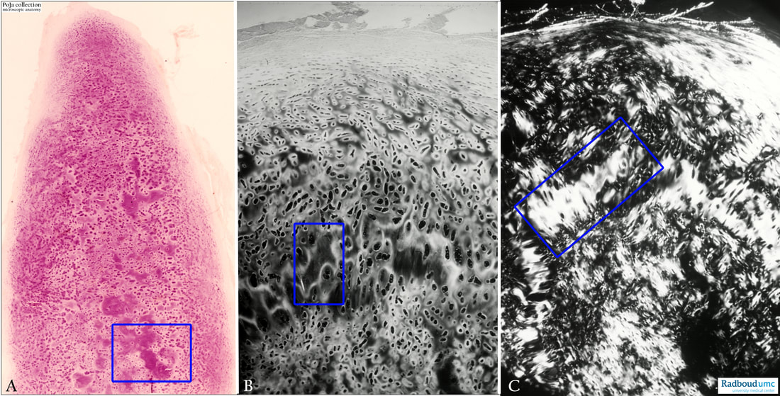
(A): Human hyaline cartilage. Haematoxylin-eosin staining. Low survey photograph. From the peripheral perichondrium to inwards, the small spindle-like chondroblasts of mesenchymal origin proliferate into islands of isogenous chondrocytes (seen as abundant small purple spots) surrounded by darkly stained extracellular matrix. Subsequently, they form irregularly larger areas which are characterised by purple stained fibrillated interterritorial matrix (rectangle). Isogenous groups of chondrocytes are clustered chondrocytes which remain together and represent recently divided cells.
(B): The matrix around the isogenous cells and between the groups of chondrocytes is stained brightly. The irregular larger areas (rectangle) are darkly stained with exception of the light boundaries (black & white image).
(C): Polarisation microscope, by changing the angle of the polarisation filters the interterritorial matrix is visualised as clear white (rectangle) due to the birefringence of collagen fibres.
Background:
The rectangles represent areas of asbestoid or amianthoid transformation (or change) formerly called asbestoid degeneration. With ageing or after damage in the cartilage, the fibrillated areas are unmasked without any presence of isogenous chondrocytes. The ongoing transformation process results in an increasing anisotropy of the collagen fibrils. The normal collagen fibrils appear to fuse together (Ghadially 2013).
See also:
Reference:
Keywords/Mesh: locomotor system, cartilage, hyaline, polarisation microscopy, matrix, collagen fibre, histology, POJA collection
Title: Hyaline cartilage visualised with polarisation microscopy
Description:
(A): Human hyaline cartilage. Haematoxylin-eosin staining. Low survey photograph. From the peripheral perichondrium to inwards, the small spindle-like chondroblasts of mesenchymal origin proliferate into islands of isogenous chondrocytes (seen as abundant small purple spots) surrounded by darkly stained extracellular matrix. Subsequently, they form irregularly larger areas which are characterised by purple stained fibrillated interterritorial matrix (rectangle). Isogenous groups of chondrocytes are clustered chondrocytes which remain together and represent recently divided cells.
(B): The matrix around the isogenous cells and between the groups of chondrocytes is stained brightly. The irregular larger areas (rectangle) are darkly stained with exception of the light boundaries (black & white image).
(C): Polarisation microscope, by changing the angle of the polarisation filters the interterritorial matrix is visualised as clear white (rectangle) due to the birefringence of collagen fibres.
Background:
The rectangles represent areas of asbestoid or amianthoid transformation (or change) formerly called asbestoid degeneration. With ageing or after damage in the cartilage, the fibrillated areas are unmasked without any presence of isogenous chondrocytes. The ongoing transformation process results in an increasing anisotropy of the collagen fibrils. The normal collagen fibrils appear to fuse together (Ghadially 2013).
See also:
- 15.0 POJA-L7006+7007+7011 Hyaline cartilage
- 15.0 POJA-L7014+7015 Hyaline cartilage with asbestos transformation (polarisation)
Reference:
- Ultrastructural pathology of the cell and matrix F.N. Ghadially 4th edition 2013, Butterworth-Heinemann ISBN-13: 978-0750698566
- Amianthoid (asbestoid) transformation: electron microscopical studies on aging human costal cartilage. R Mallinger, L Stockinger. The American Journal of Anatomy PMID: 3348145DOI: 10.1002/aja.1001810104Jan 1, 1988Paper
Keywords/Mesh: locomotor system, cartilage, hyaline, polarisation microscopy, matrix, collagen fibre, histology, POJA collection

