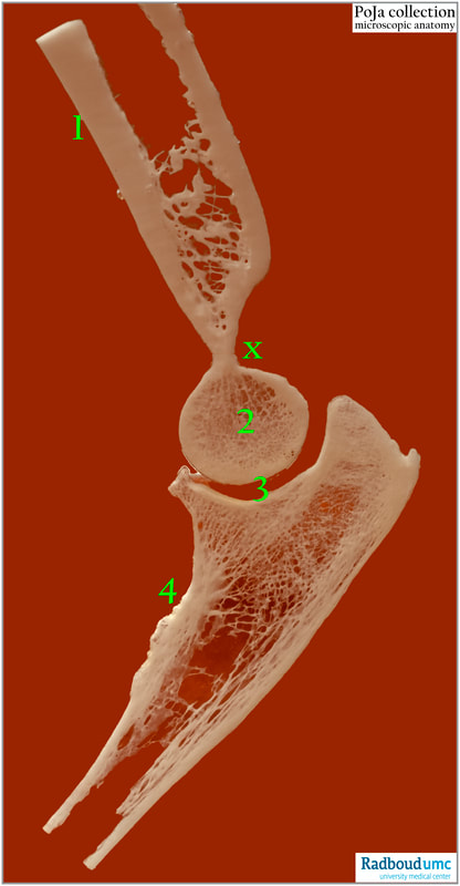
16.1.2 POJA-L7152 Anatomy of the human elbow bones
|
(By courtesy of J. Kooloos PhD and L. Boer PhD Department Medical Imaging, Anatomy, Museum for Anatomy and Pathology, Radboud university medical center, Nijmegen The Netherlands)
|
16.1.2 POJA-L7152 Anatomy of the human elbow bones
Title: Anatomy of the human elbow bones Description: The human elbow is composed of three articulations and one of them is the humero-ulnar joint (or trochlear joint). The joint is a simple hinge (the flexors and extensors use the same axis): the trochlea (2) of the humerus (1) articulates with the semilunar notch of the ulna (4): (1): Corpus humeri (2): Trochlea humeri (3): Cavum articulare (4): Ulna (x): Fossa olecrani (is a furrow however *more dorsal to the right) The bones consists of spongious trabecular bone surrounded by a shaft of compact lamellar bone. Between the trabeculae bone marrow is located containing the haematopoietic stem cells. The bone is formed by endochondral ossification process. Remember that the head of the bones in a joint is covered with cartilage. See also:
Key words/Mesh: locomotor system, bone, elbow, joint, humerus, ulna, macroscopy, histology, POJA collection |

