
13.1 POJA-L4651+La-0324+4652+3728+La-0305
Liver sinusoids
13.1 POJA-L4651+La-0324+4652+3728+La-0305
Title: Liver sinusoids
Description:
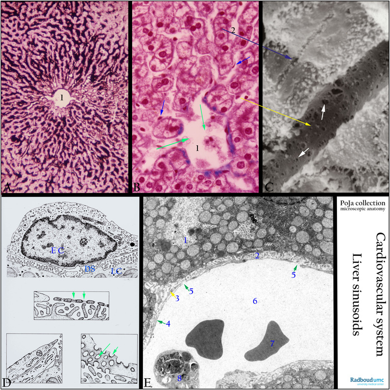
(A): Eosin staining. Liver sinusoid perfused with Indian Ink counterstained with eosin (dog). These special type of discontinuous capillaries or sinusoids forms intricate systems of anastomosing radial channels that supply individual liver cells with oxygen and nourishments. Outflow directly into central vein (1).
(B): Sinusoids (liver), Azan, human. Free influx from sinusoids into the central vein (green arrows). Endothelial cells (blue arrow) lining the sinusoids [yellow arrow, see (C)]. Bile canaliculi between hepatocytes [2, dark arrow in (C)].
(C): SEM of sinusoid with fenestrae (white arrows) of variable diameter, yellow arrow is the flattened endothelial cell (gerbil) (by courtesy of K. Theeuwes, MD).
(D): Scheme of the space of Disse lined by fenestrated endothelium cells, human. (EC) Endothelial cell. (LC) Liver cell. (DS) Space of Disse. The endothelial cell lines the sinusoid, its cytoplasm contains gaps resembling fenestrae of varying diameter (green arrows) with a ‘sieve’ function (middle scheme). Between endothelial cell and liver cells lies the space of Disse occupied by microvilli of hepatocytes and few thin collagen fibrils (type III). (Bottom left) Thin processes of the liver fibroblast (fat-storing cell) run in the space of Disse. (Bottom right) tangential section of gaps of variable diameters (green arrows). With ageing basal membrane material might be deposited.
(E): Structures identical to (D) are shown in this electron micrograph (rat). (1) Hepatocyte with microvilli in space of Disse (2). (Arrow 3) thin processes of fat-storing cell. (Arrow 4) thin cytoplasm of endothelial cell and (arrow 5) gaps. (6) Lumen of sinusoid with free erythrocytes (7) and phagocytosed material in lysosomes (8).
Background: Discontinuous capillaries or sinusoids are characterised by incomplete basal lamina and gaps (larger than fenestrae) in endothelium (liver, spleen, adrenal gland). They are endothelial-lined channels with a larger diameter than capillaries and discontinuous or even absent basement membranes.
Keywords/Mesh: cardiovascular system, vascularisation, liver, sinusoid , endothelium, gap, basal lamina, Disse space, Kupfer cells, histology, electron microscopy, POJA collection
Title: Liver sinusoids
Description:
(A): Eosin staining. Liver sinusoid perfused with Indian Ink counterstained with eosin (dog). These special type of discontinuous capillaries or sinusoids forms intricate systems of anastomosing radial channels that supply individual liver cells with oxygen and nourishments. Outflow directly into central vein (1).
(B): Sinusoids (liver), Azan, human. Free influx from sinusoids into the central vein (green arrows). Endothelial cells (blue arrow) lining the sinusoids [yellow arrow, see (C)]. Bile canaliculi between hepatocytes [2, dark arrow in (C)].
(C): SEM of sinusoid with fenestrae (white arrows) of variable diameter, yellow arrow is the flattened endothelial cell (gerbil) (by courtesy of K. Theeuwes, MD).
(D): Scheme of the space of Disse lined by fenestrated endothelium cells, human. (EC) Endothelial cell. (LC) Liver cell. (DS) Space of Disse. The endothelial cell lines the sinusoid, its cytoplasm contains gaps resembling fenestrae of varying diameter (green arrows) with a ‘sieve’ function (middle scheme). Between endothelial cell and liver cells lies the space of Disse occupied by microvilli of hepatocytes and few thin collagen fibrils (type III). (Bottom left) Thin processes of the liver fibroblast (fat-storing cell) run in the space of Disse. (Bottom right) tangential section of gaps of variable diameters (green arrows). With ageing basal membrane material might be deposited.
(E): Structures identical to (D) are shown in this electron micrograph (rat). (1) Hepatocyte with microvilli in space of Disse (2). (Arrow 3) thin processes of fat-storing cell. (Arrow 4) thin cytoplasm of endothelial cell and (arrow 5) gaps. (6) Lumen of sinusoid with free erythrocytes (7) and phagocytosed material in lysosomes (8).
Background: Discontinuous capillaries or sinusoids are characterised by incomplete basal lamina and gaps (larger than fenestrae) in endothelium (liver, spleen, adrenal gland). They are endothelial-lined channels with a larger diameter than capillaries and discontinuous or even absent basement membranes.
Keywords/Mesh: cardiovascular system, vascularisation, liver, sinusoid , endothelium, gap, basal lamina, Disse space, Kupfer cells, histology, electron microscopy, POJA collection