
11.5 POJA-L3160+4457+
3048+4458+4459.
Choroid plexus and ependym of cerebrum
11.5 POJA-L3160+4457+3048+4458+4459
Title: Choroid plexus and ependym of cerebrum
Description:
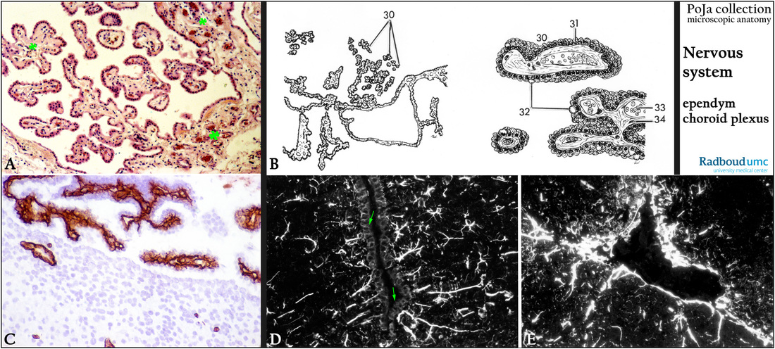
(A): Stain hematoxylin-eosin, human. Choroid plexus is a plexus in the ventricles of the brain where cerebrospinal fluid (CSF) is produced by modified ependym cells at a rate of about 20 ml per hour. The cuboidal cells, being continuous with the ventricle lining epithelium, are arranged around a core of capillaries (**).
(B): Scheme of choroid plexus, human. (30) Cross-sections of the processes. (31) Single layer of cuboidal cells.
(32) Paraplasmatic inclusions. (33) Dilated capillaries. (34) Stroma.
(C): Immunoperoxidase staining with AEC and antibodies against collagen IV, 4d postnatal rat. Collagen IV in the basement membrane lines the cuboidal cells and is also present in the stroma of processes of the choroid plexus. Note that a basement membrane is missing below the normal ependymal lining of the ventricle!!
(D): Immunofluorescence staining with antibodies against GFAP (glial fibrillary acidic protein), black-white, mouse. Astrocytes are positive. However, note that the normal lining ependym (arrows) of the 3rd ventricle is negative.
(E): Immunofluorescence staining with antibodies against vimentin, black-white, mouse. Most of the vimentin-fluorescence is located in the subependymal lining of a gyrus, within the perivascular spaces and less in the astrocytes.
Keywords/Mesh: nervous tissue, cerebrum, ventricle, ependym, choroid plexus, cerebrospinal fluid, GFAP, vimentin, histology, POJA collection
Title: Choroid plexus and ependym of cerebrum
Description:
(A): Stain hematoxylin-eosin, human. Choroid plexus is a plexus in the ventricles of the brain where cerebrospinal fluid (CSF) is produced by modified ependym cells at a rate of about 20 ml per hour. The cuboidal cells, being continuous with the ventricle lining epithelium, are arranged around a core of capillaries (**).
(B): Scheme of choroid plexus, human. (30) Cross-sections of the processes. (31) Single layer of cuboidal cells.
(32) Paraplasmatic inclusions. (33) Dilated capillaries. (34) Stroma.
(C): Immunoperoxidase staining with AEC and antibodies against collagen IV, 4d postnatal rat. Collagen IV in the basement membrane lines the cuboidal cells and is also present in the stroma of processes of the choroid plexus. Note that a basement membrane is missing below the normal ependymal lining of the ventricle!!
(D): Immunofluorescence staining with antibodies against GFAP (glial fibrillary acidic protein), black-white, mouse. Astrocytes are positive. However, note that the normal lining ependym (arrows) of the 3rd ventricle is negative.
(E): Immunofluorescence staining with antibodies against vimentin, black-white, mouse. Most of the vimentin-fluorescence is located in the subependymal lining of a gyrus, within the perivascular spaces and less in the astrocytes.
Keywords/Mesh: nervous tissue, cerebrum, ventricle, ependym, choroid plexus, cerebrospinal fluid, GFAP, vimentin, histology, POJA collection