
16.1.2 POJA-L7151 Anatomy of the knee joint
16.1.2 POJA-L7151 Anatomy of the knee joint
(By courtesy of J. Kooloos PhD and L. Boer PhD, Department Medical Imaging, Anatomy, Museum for Anatomy and Pathology, Radboud university medical center, Nijmegen The Netherlands)
16.1.2 POJA-L7151 Anatomy of the knee joint
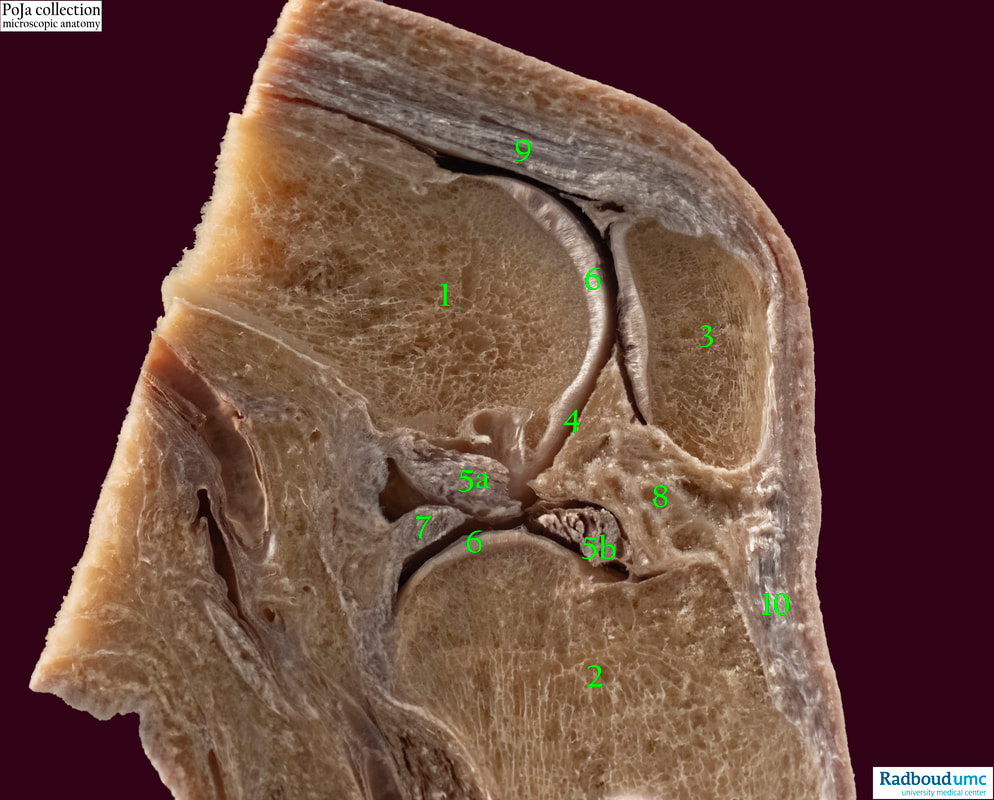
Title: Anatomy of the knee joint
Description:
Sagittal section of knee joint, human:
(1): Femur. (2): Tibia. (3): Patella. (4): Synovial membrane. (5a): Posterior cruciate ligament. (5b): Anterior cruciate ligament.
(6): Articular surface. Medial side of the medial tibial plateau. (7): Medial meniscus. (8): Infrapatellar fat pad (Hoffa).
(9): Quadriceps femoris muscle. (10): Patellar ligament.
The knee comprises two separate joints, i.e., the femoropatellar (1-3) which is a sellar joint and the femorotibial (1-2) which is a bicondylar joint. The latter is further partly divided by menisci between the corresponding articular surfaces (6).
The bones (1, 2, 3) in the knee are formed through the process of enchondral ossification resulting in spongious trabecular bone surrounded by a shaft of compact lamellar bone.
The menisci show fibrocartilage (outer two-thirds) and hyaline cartilage (inner one-third).
The synovium consists of 1-4 cell layers enforced by fibrocollagenous capsule. The epithelial cell population contains cells that produce proteins for the synovial fluid, and phagocytic cell types.
The articular surface (6) is covered with cartilage.
See also:
Key words/Mesh: locomotor system, bone, cartilage, knee joint, synovium, anatomy, POJA collection, histology, femur, tibia, patella
16.1.2 POJA-L7151 Anatomy of the knee joint
Title: Anatomy of the knee joint
Description:
Sagittal section of knee joint, human:
(1): Femur. (2): Tibia. (3): Patella. (4): Synovial membrane. (5a): Posterior cruciate ligament. (5b): Anterior cruciate ligament.
(6): Articular surface. Medial side of the medial tibial plateau. (7): Medial meniscus. (8): Infrapatellar fat pad (Hoffa).
(9): Quadriceps femoris muscle. (10): Patellar ligament.
The knee comprises two separate joints, i.e., the femoropatellar (1-3) which is a sellar joint and the femorotibial (1-2) which is a bicondylar joint. The latter is further partly divided by menisci between the corresponding articular surfaces (6).
The bones (1, 2, 3) in the knee are formed through the process of enchondral ossification resulting in spongious trabecular bone surrounded by a shaft of compact lamellar bone.
The menisci show fibrocartilage (outer two-thirds) and hyaline cartilage (inner one-third).
The synovium consists of 1-4 cell layers enforced by fibrocollagenous capsule. The epithelial cell population contains cells that produce proteins for the synovial fluid, and phagocytic cell types.
The articular surface (6) is covered with cartilage.
See also:
- 16.1.4 POJA-L7127+7129 Foetal knee joint 4
- 16.1.4 POJA-L7128+7131+7133 Foetal knee joint 3
- 16.1.4 POJA-L7130+7132 Adult knee joint
Key words/Mesh: locomotor system, bone, cartilage, knee joint, synovium, anatomy, POJA collection, histology, femur, tibia, patella