10.2 POJA-L2129+2004+2220+2245
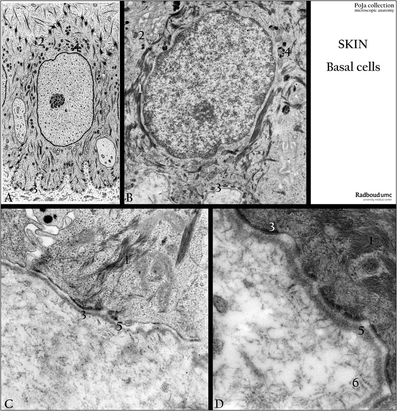
Title: Basal cell in the epidermis
Description:
(A): Electron microscopy scheme, human.
(B-D): Back skin, electron microscopy, human.
Cells in the stratum basale (or stratum germinativum) show in the cytoplasms:
(1) Tonofilaments or cytokeratin filaments.
(2) Desmosomal junctions.
(3) Hemidesmosomal junctions.
(4) Melanin granules.
(5) Basal lamina.
(6) Collagen VII fibrils with periodicity.
(Electron micrograph (B) by courtesy of A. Stadhouders, PhD, former Head Department of Electron Microscopy,
Radboud university medical center, Nijmegen, The Netherlands).
Keywords/Mesh: skin, epidermis, stratum basale, stratum germinativum, basal cell, melanin, cytokeratin, desmosome, hemidesmosome, basal lamina, collagen VII, histology, electron microscopy, POJA collection

