6.6 POJA-L2705+2707+2708
Title: Tail defects of spermatozoa in immotile cilia-syndrome
Description:
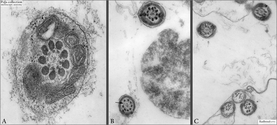
Electron micrographs of cross-sections through the tail of spermatozoa in ejaculates
(immotile-cilia syndrome), human.
(A): Tail of a normal spermatozoon (partly tangentially cut). The middle piece of the tail contains a core of an axoneme (see kinocilia in Respiratory system) i.e. 2 single central microtubules and a cylinder of 9 double peripheral microtubules. Additionally the axoneme is surrounded by 9 longitudinally orientated coarse outer fibrils. Circular mitochondria are found around this internal structure that is responsible for the contraction and the movement of the spermatozoa in a forward motion.
(B, C): Abnormal tails of possible immotile spermatozoa. Cross-sections through the principal pieces (at this level an additional continuous fibrous sheath encircles the coarse outer longitudinal bundles); note especially in B (arrows) a lack of one peripheral doublet of microtubules.
(C): Disorganization of the array of microtubules and coarse fibrils is apparent.
Keywords/Mesh: testis , sperm, spermatozoon, immotile-cilia syndrome, Ciliary Motility Disorder, histology. electron microscopy, POJA collection

