5.4.1 POJA-L2377+2369+5039+4276+4278+4277
Title: Foot processes of podocytes and glomerular basal lamina (XIII) in the kidney
Description:
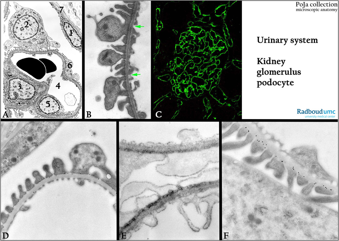
(A): Glomerulus, scheme, human. Parietal cell (1) lining the Bowman’s capsule (7). Podocyte (2) with primary and secondary
pedicles (6) around the capillary (4) lined with endothelial cells (5). Mesangium cell (3).
(B): Glomerulus, electron microscopy, rat. Glomerular basal lamina (GBL) with pedicles of the podocyte at one side and fenestrated endothelial cell cytoplasm (arrows) at the other side. The GBL is composed of three layers: as dense layer squeezed between light
outer and inner layers: close to the podocyte an electron-light outer lamina rara externa (LRE), a lamina densa (LD) and an electron-
light lamina rara interna (LRI) close to the fenestrated endothelial cytoplasm.
(C): Glomerulus, immunofluorescence staining with antibodies (4C3) against heparan sulfate showing the localization in the GBL of
the capillaries, rat.
(C, by courtesy of G.B. ten Dam, PhD, Department of Biochemistry, University Medical Centre of the Radboud University, Nijmegen,
The Netherlands).
(D, F): Glomerulus, electron microscopy, immunogold labeling with antibodies (4C3) against heparan sulfate illustrated by the 10 nm gold granules in the
basal lamina, rat. The labeling is restricted to a small zone (external lamina rara) close to the end processes of the podocytes.
(E): Glomerulus, electron microscopy, staining of the glycocalyx with antibodies (4C3) against heparan sulfate with DAB and ruthenium red, rat.
Two capillaries engulfed by one common podocyte, note the denser-stained external lamina rara of the GBL close to the feet processes. Ruthenium red also adheres as a thin fluffy lining along all cell membranes of cytoplasm and processes.
Background: HSPG as component of the basement membrane can bind to cytokines, growth factors and morphogens, creating
a depot of regulatory factors that can be liberated by selective degradation of the HSPG.
They can act as receptors for proteases/inhibitors.
They also function in combination with integrins to participate in cell-ECM (extracellular matrix) attachment and cell-cell interactions.
Keywords/Mesh: urinary system, kidney, glomerulus, glomerular basement lamina, podocyte, pedicle, capillary, fenestrated endothelium, heparan sulfate, histology, electron microscopy, POJA collection

