5.4.2 POJA-L2330+L4284+La0084
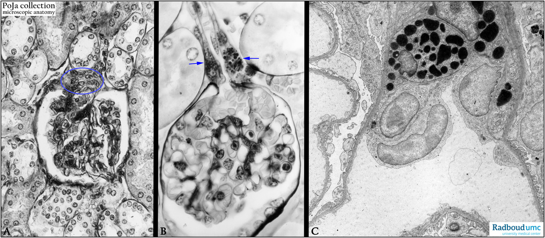
Title: Renin-containing cells in the afferent arteriole of the renal corpuscle in kidney VI
Description:
(A): Glomerulus, stain Azan (black-white), human. The encircled area indicates the afferent arteriole and the macula densa area.
(B): Glomerulus, silver stain (Movat, black-white), mouse. The granulated JG cells (arrows) with renin are located in the wall of the
vas afferens just before entering the glomerulus.
(C): Glomerulus. electron microscopy, perfusion-fixed, rat. This shows the ultrastructural equivalent of the renin granules in the
bivalent (modified smooth muscle cells) JG-cells, located just below the endothelial cells.
Keywords/Mesh: urinary system, kidney, glomerulus, vas afferens, JG cells, renin, histology, electron microscopy, POA collection

