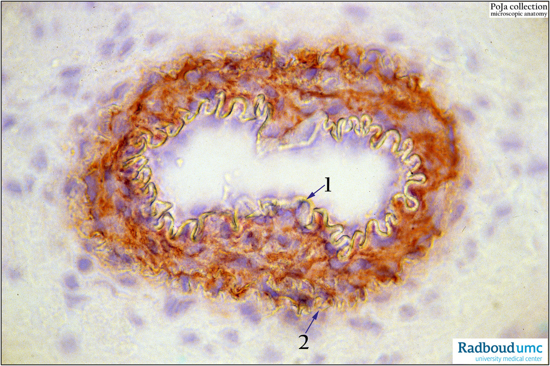
13.1 POJA-La0298
Title: Muscular media: vascular smooth muscle cells (VSM)
Description:
Small artery (inner ear region), actin, rat postnatal. Smooth muscle cells in the media of this small artery demonstrate the presence of cytoplasmic actin. Antibodies against smooth muscle actin (alpha-Sm actin) are used with AEC staining (red-brown) and nuclei counterstained with haematoxylin (frozen section). Note the translucent wavy internal elastic lamina (arrow 1) as well as the external elastic lamina (arrow 2) of this type of muscular artery.
Background: Smooth muscle cells (SMCs) in the media also referred as vascular smooth muscle (VSM): Slow and tonic contractions are characteristic for vascular smooth cells (VSM) in contrast to rapid and short duration of the contractions of cardiac muscle. The contractile proteins as actin and myosin are specifically organised and well-suited for maintaining tonic contractions and reducing the diameter of the blood vessel lumen. An increased influx of calcium leads to free calcium that binds to the calcium-binding protein calmodulin. The enzyme myosin light chain kinase (MLCK) will be activated and this process is followed by cross-bridge formation between myosin heads and actin filaments resulting in VCM contraction. VSM relaxation occurs when free intracellular calcium decreases. The presence of smooth muscle actin in the VSM is demonstrated by immunohistochemistry using monoclonal antibodies against alpha-Sm actin (ACE staining technique).
Keywords/Mesh: cardiovascular system, vascularisation, blood vessel, muscular artery, vascular smooth muscle, smooth muscle cell, elastic membrane, actin, histology, POJA collection

