10.3 POJA-L2217+2218+2222+2219
Title: Dermis of the skin
Description:
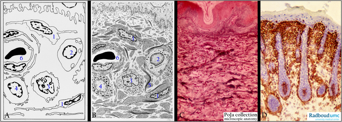
(A, B): Electron microscopy scheme of the dermis, human.
(1) Fibroblasts. (4) Mast cell.
(2) Schwann cell with axons. (5) Cross-section/longitudinal section of collagen fibers.
(3) Macrophage. (6) Capillary with pericyte.
(C): Arm skin, stain orcein-light green, human. The dermis contains different types of collagen fibers (greenish-stained)
as well as reddish-stained elastin.
(D): Back skin, immunoperoxidase staining with DAB and monoclonal antibodies (RV202) against vimentin, rat.
Mesenchyme-derived cells (dermis) are stained positively but not the epithelial cell types (keratinocytes).
Note the presence of hair follicles.
Keywords/Mesh: skin, dermis, vimentin, collagen, elastin, histology, electron microscopy, POJA collection

