
2.1 POJA-L947. Immunohistochemistry of ER13, ED1, ED2, ED3, ER2 markers in normal thymus (rat)
2.1 POJA-L947
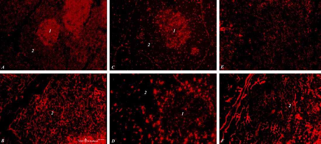
Title: Immunohistochemistry of ER13, ED1, ED2, ED3, ER2 markers in normal thymus (rat)
Description: Stain: Alexa-594 red immunofluorescence.
(1): Medulla.
(2): Cortex of the thymus.
(A-survey, B-cortex): ER13 antibody stains for MHC-class II antigens on reticular cell types in medulla and cortex.
(C): ED1 monoclonal antibody stains a single chain glycoprotein of 110 kDa on the lysosomal membrane of myeloid cells, i.e. the majority of tissue macrophages (being the rat homologue of human CD68);
(D): ED2 monoclonal antibody reacts with a membrane antigen (175, 160 and 95 kDa) on resident rat macrophages such as monocytes, dendritic cells. ED2 discriminates between thymic cortical (positive for ED2) and medullary (negative for ED2) macrophages. The antigen is identical with CD163.
(E): ER2 antibody stains the CD4 rat antigen more intensely expressed on mature, medullary thymocytes.
(F): ED3 monoclonal antibody recognises the rat CD169 cell surface antigen, a 185 kDa molecule expressed by macrophages in lymphoid organs (no monocytes or granulocytes). In the thymus the antigen is expressed on clusters of dendritic cells (thymic nurse cells or TNC’s) in the (outer) cortex.
Keywords/Mesh: lymphatic organs, thymus, immunohistochemistry, ER2, ER13, MHC-II, ED1, ED2, ED3, rat, toxicology, histology, POJA collection