
1.1 POJA-L774. Mast cell (connective tissue, human)
|
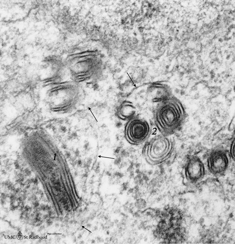
1.1 POJA-L774
Title: Mast cell (connective tissue, human) Description: Electron microscopy. Mast cells (mastocytes) are frequently found perivascularly or perineurally within the connective tissue. A detail of this mast cell shows granules varying in shape and size. These membrane-bound vesicles (so-called compound granules) show a metachromatic reaction in light microscopy and ultrastructurally a granule exhibits a heterogeneous content (different with species). The internal substructure is composed of osmiophilic short parallel stalks (1) in a granular substance appearing in cross-sections as scroll-like or whorl-like figures (2). Scroll-like as well as whorl-like substructures are common in human mastocytes. At thin arrows (↓) the boundaries of some granules. These granules contain among others heparin, histamine, enzymes such as superoxide dismutase, b-hexosaminidase, tryptase, factors such as neutrophil – and eosinophil-chemotactic factors, vasoactive mediators. |
Background:
Analogous to basophilic granulocytes mast cells possess specific membrane receptors for the Fc segment of IgE produced in response to allergens. Eventually the release of the granular content by exocytosis (leading to degranulation of the mast cell) occurs resulting in e.g. an immediate hypersensitivity (anaphylactoid) reaction.
Keywords/Mesh: blood, bone marrow, basophilic granulocyte, specific granule, mast cell, histamine, histology, electron microscopy, POJA collection