
10.2 POJA-L2138+2139+2140
+2141+2142+2143.
Lamellated granules (Odland bodies) in stratum granulosum of the epidermis
10.2 POJA-L2138+2139+2140+2141+2142+2143
Title: Lamellated granules (Odland bodies) in stratum granulosum of the epidermis
Description:
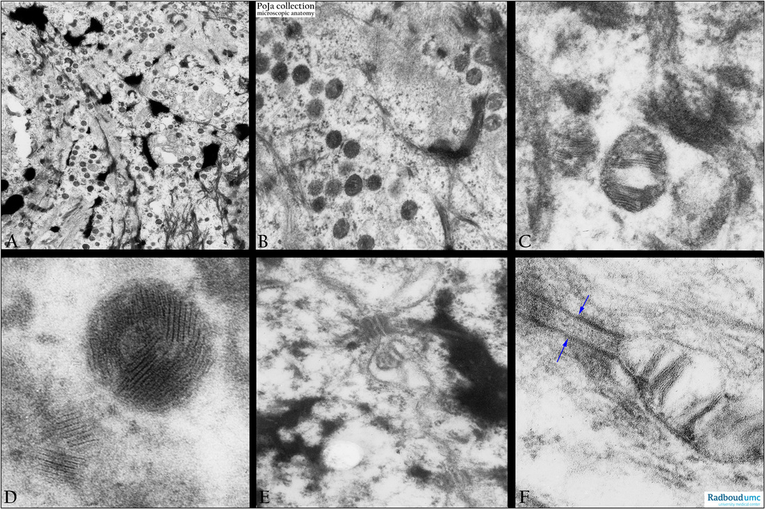
Back skin, electron microscopy, human.
(A, B): Stratum granulosum with electron-dense keratohyalin aggregates, detailed in (C- D -F).
(C-D:) Membrane-coating granules (or lamellar granules, lamellated granules, Odland bodies) with internal lamellae.
(E): Intercellular localized Odland body close to a desmosome.
(F): Higher magnification of an intercellular Odland body. (Arrows) two apposed cell membranes of adjacent cells.
Odland bodies contain glycolipidacylglucosylceramid which is extruded in the intercellular spaces between the cells of
the granular layer (stratum granulosum) for coating of the corneocytes, thus providing a water barrier (“brick and mortar” model).
(Electron micrographs (D, F) by courtesy of A. Stadhouders, PhD, Head of the former Department of Electron Microscopy,
Radboud university medical center, Nijmegen, The Netherlands).
Keywords/Mesh: skin, epidermis, stratum granulosum, granular layer, keratohyalin, Odland body, membrane-coating granule,
lamellated granule, histology, electron microscopy, POJA collection
Title: Lamellated granules (Odland bodies) in stratum granulosum of the epidermis
Description:
Back skin, electron microscopy, human.
(A, B): Stratum granulosum with electron-dense keratohyalin aggregates, detailed in (C- D -F).
(C-D:) Membrane-coating granules (or lamellar granules, lamellated granules, Odland bodies) with internal lamellae.
(E): Intercellular localized Odland body close to a desmosome.
(F): Higher magnification of an intercellular Odland body. (Arrows) two apposed cell membranes of adjacent cells.
Odland bodies contain glycolipidacylglucosylceramid which is extruded in the intercellular spaces between the cells of
the granular layer (stratum granulosum) for coating of the corneocytes, thus providing a water barrier (“brick and mortar” model).
(Electron micrographs (D, F) by courtesy of A. Stadhouders, PhD, Head of the former Department of Electron Microscopy,
Radboud university medical center, Nijmegen, The Netherlands).
Keywords/Mesh: skin, epidermis, stratum granulosum, granular layer, keratohyalin, Odland body, membrane-coating granule,
lamellated granule, histology, electron microscopy, POJA collection