
13.1 POJA-La0284+L4683+4562+4684+4565+4577 Media of elastic arteries
13.1 POJA-La0284+L4683+4562+4684+4565+4577
Title: Media of elastic arteries
Description:
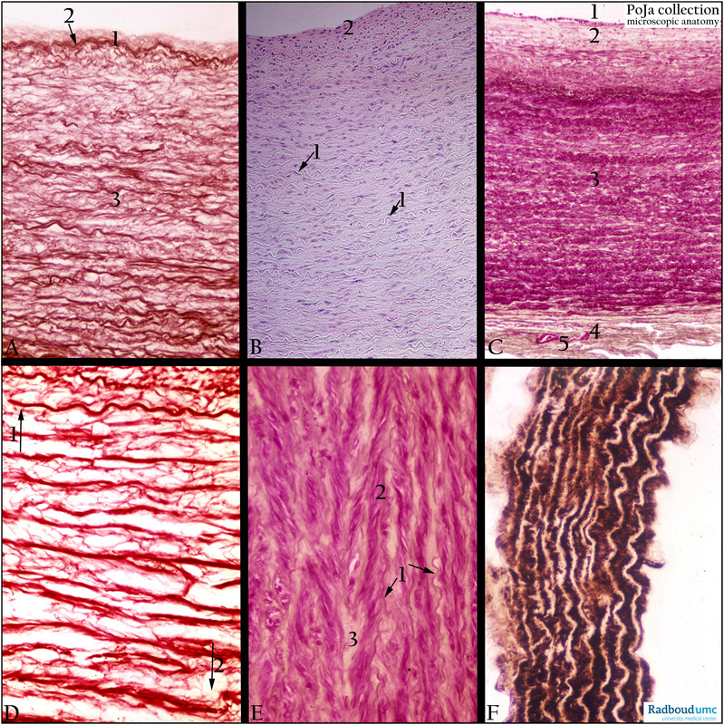
(A): Carotid artery, orcein, human. Intima (1) and at (arrow 2) the first thick elastic lamella followed by numerous elastic lamellae or membranes in the broad and well developed media (3).
(B): Common carotid artery, HE, human. When contrast is enhanced by closing the microscope diaphragm aperture numerous unstained elastic membranes in the media are visualised as tortuous unstained transparent laminae (arrow 1). Intima (2).
(C): Aorta, resorcin-fuchsin and light green, human. Lumen of aorta (1), intima ca. 150 micrometers (2), thick media ca. 2 mm (3), thin adventitia (4) with little vasa vasorum (5). The latter comprises small vessels penetrating the outer part of the media for the supply of oxygen. The media contains about 50-75 concentric elastic fenestrated membranes (ca. 2-5 micrometers) with interlinking branched smooth muscle cells (SMC).
(D): Aorta, orcein, human. Direction of intima (arrow 1) and (arrow 2) direction of adventitia. Only dense-stained elastic membranes in the media, between the membranes thin branches of elastic fibres envelop SMCs (not stained, not visible).
(E): Media of aorta, resorcin-fuchsin and modified light green, human. A part of the aortic media shows the undulant elastic membranes (arrow, 1) while branched smooth muscle cells (SMC) (2) embedded in light-brown extracellular matrix (ECM) (3) are interlinked between elastic membranes in a so-called herringbone pattern.
(F): Mg-ATPase staining on the aorta smooth muscle cells (SMC), mouse. Note that the SMC are strongly positive while the elastic membranes remain negative.
Background: Elastic arteries exhibit media with concentric elastic fenestrated membranes with interlinking branched smooth muscle cells (SMC). Due to the presence of these elastic laminae as well as that of the medial SMCs these so-called conducting arteries are resistant to systolic pressure. As viscoelastic vessels they act like an air chamber (“Windkessel” effect) and propagate heart contraction and smoothing out the pulsatile flow. In this way they support a continuous blood circulation despite the intermittent pumping heart. Examples of these arteries are aorta, brachiocephalic trunk, common carotid artery, and subclavian artery, and thyrocervical trunk, proximal parts of the vertebral artery, extrapulmonary artery, internal thoracic artery, and common iliac artery. Many larger arteries e.g. common carotid artery possess less elastic membranes in the media but more in the adventitia. The higher amount of SMCs in the media also contribute to the stiffness of this artery.
Keywords/Mesh: cardiovascular syatem, vascularisation, blood vessel, elastic artery , elastic lamella, vasa vasorum, aorta, carotid artery, histology, POJA collection
Title: Media of elastic arteries
Description:
(A): Carotid artery, orcein, human. Intima (1) and at (arrow 2) the first thick elastic lamella followed by numerous elastic lamellae or membranes in the broad and well developed media (3).
(B): Common carotid artery, HE, human. When contrast is enhanced by closing the microscope diaphragm aperture numerous unstained elastic membranes in the media are visualised as tortuous unstained transparent laminae (arrow 1). Intima (2).
(C): Aorta, resorcin-fuchsin and light green, human. Lumen of aorta (1), intima ca. 150 micrometers (2), thick media ca. 2 mm (3), thin adventitia (4) with little vasa vasorum (5). The latter comprises small vessels penetrating the outer part of the media for the supply of oxygen. The media contains about 50-75 concentric elastic fenestrated membranes (ca. 2-5 micrometers) with interlinking branched smooth muscle cells (SMC).
(D): Aorta, orcein, human. Direction of intima (arrow 1) and (arrow 2) direction of adventitia. Only dense-stained elastic membranes in the media, between the membranes thin branches of elastic fibres envelop SMCs (not stained, not visible).
(E): Media of aorta, resorcin-fuchsin and modified light green, human. A part of the aortic media shows the undulant elastic membranes (arrow, 1) while branched smooth muscle cells (SMC) (2) embedded in light-brown extracellular matrix (ECM) (3) are interlinked between elastic membranes in a so-called herringbone pattern.
(F): Mg-ATPase staining on the aorta smooth muscle cells (SMC), mouse. Note that the SMC are strongly positive while the elastic membranes remain negative.
Background: Elastic arteries exhibit media with concentric elastic fenestrated membranes with interlinking branched smooth muscle cells (SMC). Due to the presence of these elastic laminae as well as that of the medial SMCs these so-called conducting arteries are resistant to systolic pressure. As viscoelastic vessels they act like an air chamber (“Windkessel” effect) and propagate heart contraction and smoothing out the pulsatile flow. In this way they support a continuous blood circulation despite the intermittent pumping heart. Examples of these arteries are aorta, brachiocephalic trunk, common carotid artery, and subclavian artery, and thyrocervical trunk, proximal parts of the vertebral artery, extrapulmonary artery, internal thoracic artery, and common iliac artery. Many larger arteries e.g. common carotid artery possess less elastic membranes in the media but more in the adventitia. The higher amount of SMCs in the media also contribute to the stiffness of this artery.
Keywords/Mesh: cardiovascular syatem, vascularisation, blood vessel, elastic artery , elastic lamella, vasa vasorum, aorta, carotid artery, histology, POJA collection