
12.2.4 POJA-L2629+La0104+
La-0099+2613+2631
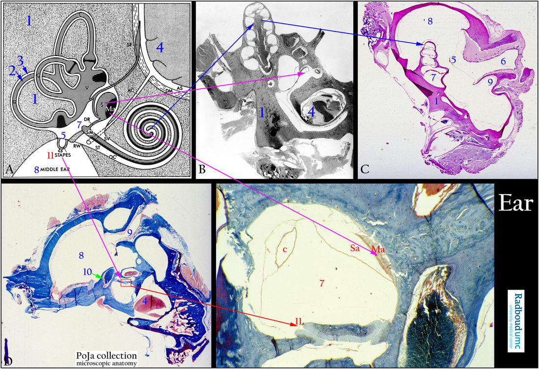
Inner ear with organ of Corti
12.2.4 POJA-L2629+La0104+La0099+2613+2631
Title: Inner ear with organ of Corti
Description:
(A): Scheme, human. (B): Stain Azan (black and white print), guinea pig. (C): Stain Haematoxylin-eosin, middle ear (bulla, tympanic cavity), guinea pig.
(D): Stain Azan, bulla with stapes, guinea pig. (E): Stain Azan, macula/sacculus, semicircular canal, foot plate, stapedius muscle, guinea pig.
(A): Scheme inner ear. (By courtesy of W. Kuipers, PhD, former Head Biological Laboratory, Department of Otorhinolaryngology,
Radboud university medical center, Nijmegen, The Netherlands).
In the osseous labyrinth (A, 1; B, 1; C, 1; D, 1) lies the membranous labyrinth (A, 2) and between them a space (3) filled with delicate connective tissue and capillaries the so-called perilymphatic tissue where perilymph circulates.
The membranous labyrinth contains the auditory (cochlea) and vestibular system (semicircular canals).
(A, 4) Cerebrum. (D, 10) Stapedius muscle.
(A, 5; C, 5) Tympanic membrane. (A, 11; D, 11; E, 11) Stapes.
(C, 6) External meatus. (E, 7) Vestibulum.
(A, 7; C, 7) Vestibulum. (E) Sa is sacculus as shown in (A, S).
(C, 9) Begin of Eustachian tube. (E) Ma is macula in the saculus also shown in (A, Ma).
(A, 8; C, 8; D, 8) Middle ear or bulla tympani.
(SV) Scala vestibuli. (RW) Round window.
(ST) Scala tympani. (AS) Subarachnoid space.
(OC) Organ of Corti. (DM) Dura mater and subdural space.
(AC) Cochlear aqueduct (or perilymphatic duct) from the subarachnoidal space to scala tympani
(perilymph is analogue to cerebrospinal liquor).
The endolymph in the scala media (membranous cochlear duct) (SM) runs via the ductus reuniens (DR) to the saccule (S)
and from there into the endolympatic duct to the endolymphatic sac (SE) within the subdural space.
(C) The so-called three semicircular arches are situated in three different planes.
These semicircular canals are involved in angular acceleration i.e. a change in the rate of head rotation.
The semicircular canals open into a portion of the membranous labyrinth, the so-called utricle (U),
the endolymph runs into the endolymphatic sac (SE).
Keywords/Mesh: inner ear, cochlea, cochlear duct, vestibulum, vestibular organ, histology, POJA collection