
12.2.4.2 POJA-L3417+3420+
3407+3410
Macula of the sacculus in the inner ear
12.2.4.2 POJA-L3417+3420+3407+3410
Title: Macula of the sacculus in the inner ear
Description:
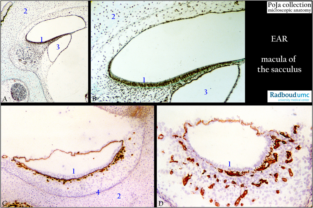
(A, B): Development of the macula of the saccule, stain trichrome Goldner, fetus human. The future macula (1) is a small patch of maturing sensory epithelium without otoconia (or statoconia) attached to the perichondrium of the hyaline cartilaginous labyrinth (2) (in adulthood the future endosteum of the osseus labyrinth). Adjacent to it a cross-section of part of a semicircular arcade (3).
(C, D): Macula, immunoperoxidase staining with AEC and antibodies against collagen IV, 1 day postnatal rat. The position of blood vessels and punctuated nerve fibres just below the sensory epithelial cells are well marked by basal lamina containing collagen IV.
Cross-section of the saccule is positively lined by collagen IV in the basal lamina. Between the membranous canal and the cartilaginous labyrinth a connective tissue network collagen IV-positive blood vessels are present (perilymphatic tissue). In (D) another area at higher magnification demonstrates the rich vascularisation. The hyaline cartilaginous tube is surrounded by a layer of denser–stained perichondrial cells (4) that give rise to the chondroblasts (in C). Postmitotic daughter cells remain together. Between the cell groups
the interritorial matrix stains weakly bluish. During maturing the hyaline cartilage will be replaced by (see 15.1 POJA-L7006+7007+7011 Hyaline cartilage; 16.1.3 POJA-L7109+7108+7101+7097 Endochondral ossification in foetus 4).
Keywords/Mesh: inner ear, fetus, semicircular canal, saccule, macula, collagen IV, histology, POJA collection
Title: Macula of the sacculus in the inner ear
Description:
(A, B): Development of the macula of the saccule, stain trichrome Goldner, fetus human. The future macula (1) is a small patch of maturing sensory epithelium without otoconia (or statoconia) attached to the perichondrium of the hyaline cartilaginous labyrinth (2) (in adulthood the future endosteum of the osseus labyrinth). Adjacent to it a cross-section of part of a semicircular arcade (3).
(C, D): Macula, immunoperoxidase staining with AEC and antibodies against collagen IV, 1 day postnatal rat. The position of blood vessels and punctuated nerve fibres just below the sensory epithelial cells are well marked by basal lamina containing collagen IV.
Cross-section of the saccule is positively lined by collagen IV in the basal lamina. Between the membranous canal and the cartilaginous labyrinth a connective tissue network collagen IV-positive blood vessels are present (perilymphatic tissue). In (D) another area at higher magnification demonstrates the rich vascularisation. The hyaline cartilaginous tube is surrounded by a layer of denser–stained perichondrial cells (4) that give rise to the chondroblasts (in C). Postmitotic daughter cells remain together. Between the cell groups
the interritorial matrix stains weakly bluish. During maturing the hyaline cartilage will be replaced by (see 15.1 POJA-L7006+7007+7011 Hyaline cartilage; 16.1.3 POJA-L7109+7108+7101+7097 Endochondral ossification in foetus 4).
Keywords/Mesh: inner ear, fetus, semicircular canal, saccule, macula, collagen IV, histology, POJA collection