|
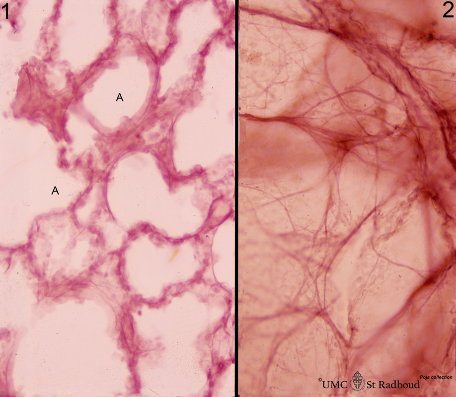
8.5 POJA-L398 Title: Elastic fibers in the lung Description: Stain orcein, human. At the left (1) several alveoli (A) are depicted with a faint staining of cellular elements. The elastic fibers within the alveoli are stained reddish-purple. At the right (2) a high magnification of a tangential-sectioned alveolus shows the thicker bundles as well as very thin branches of elastic fibers. |
|
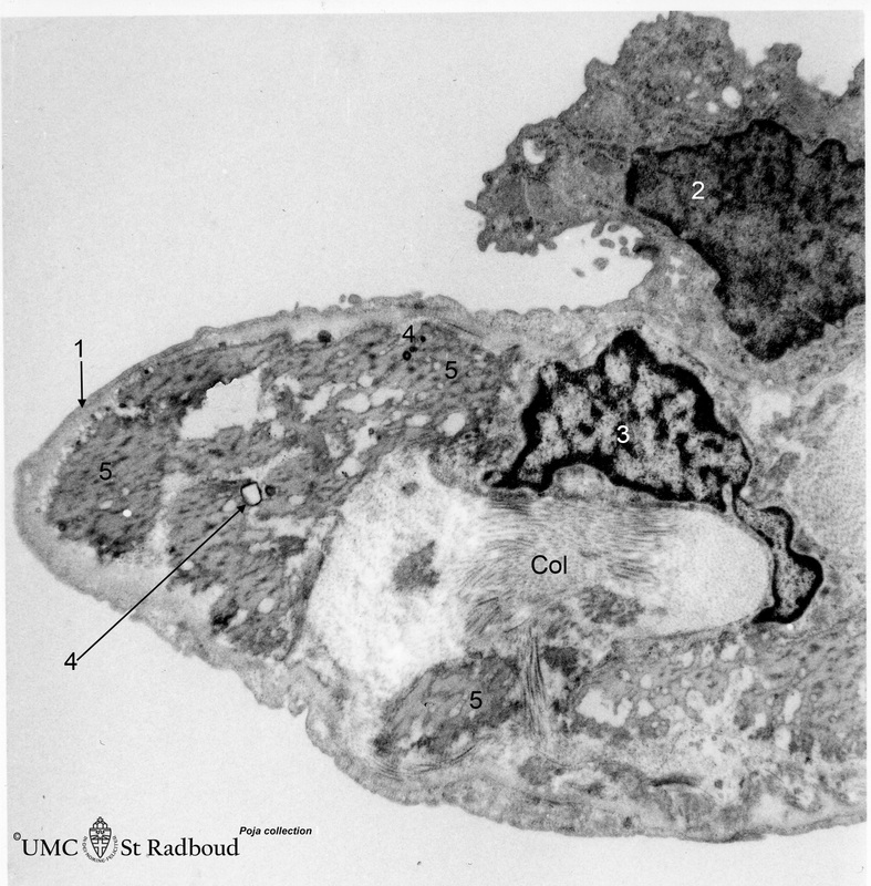
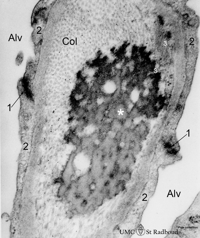
8.5 POJA-L464 Title: The alveolar tip in lung tissue Description: Electron microscopy, adult human. The lung tip is covered by a flattened alveolar cell type I (↓1) and a similar neighbouring bulging one with nucleus (2). In the alveolar tip amorph elastin (5) appears more electron-dense than the collagen fibers (Col) and is interwoven with it. Small foci of calcifications are found common in ageing lungs (4); (3) indicates an elastoblast in close association with the surrounding elastin as well as with bundles of collagen (Col). 8.5 POJA-L466 Title: Elastin close near the alveolar tip in lung tissue Description: Electron microscopy, adult human. The alveolar tip in the alveolar spaces (Alv) is covered with flattened type I alveolar cells (2). The arrows at (↓1) indicate the junctions between these flattened cells. The amorph elastin (*) in the alveolar tip appears more electron-dense than the collagen fibers (Col). Note the characteristic dark dots and bundles of microfibrils (3) (among others fibrillin) associated with elastin. Keywords/Mesh: respiratory system, lung, alveolus, alveolar tip, type I alveolar cell, pneumocyte I, myofibroblast, elastoblast, collagen, elastic fiber, elastin, microfibril, elastin-associated protein, histology, electron microscopy, POJA collection |


