4.2.1 POJA-L2931-2932
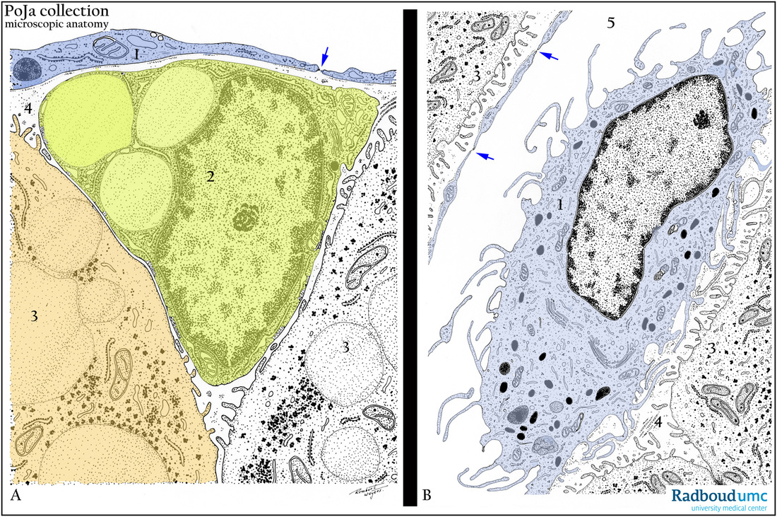
Title: Scheme of sinusoid endothelial cells and hepatic stellate cell (human)
Description: Electron micrograph scheme of (A) Hepatic stellate cell (cell of Ito) (2, yellow color), and (B) part of a sinusoid endothelial cell and Kupffer cell both located in the sinusoid space of the liver (5). The hepatic stellate cell is located within the space of Disse (4), i.e. between the lining endothelial cell (1, blue color) and the liver cell (3) that bears microvilli. The sinusoid endothelial has long slender extensions resting just above the microvilli of the liver cell. Arrows (↘) are fenestrations in the thin cell processes. The Kupffer cell (in B) is characterized by many lysosomes, Golgi areas and numerous thin filopodia.
Keywords/Mesh: liver cell, hepatic stellate cell, sinusoid endothelial cell, Kupffer cell, sinusoid, histology, electron microscopy, POJA collection

