
14.6.1 POJA-L6190B+6188+6189 Mitochondrial myopathy I: Ragged-red fibres (human)
14.6.1 POJA-L6190B+6188+6189 Mitochondrial myopathy I
14.6.1 POJA-L6190B+6188+6189 Mitochondrial myopathy I: Ragged-red fibres (human)
(Micrographs by courtesy of H. ter Laak PhD Section Neuropathology, retired staff member Department of Pathology, Radboud university medical center, Nijmegen, The Netherlands)
Title: Mitochondrial myopathy I: Ragged-red fibres (human)
Description:
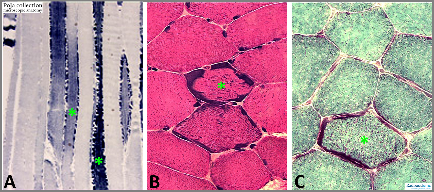
(A): SDH (succinate dehydrogenase) staining of ragged-red muscle fibres. (B): Haematoxylin eosin staining of ragged-red muscle fibres. (C): Trichrome staining. Asterix indicate the ragged-red fibres.
Background:
These ragged-red fibres appear red in the Trichrome stain. The “ragged” appearance is caused by the mitochondrial aggregation just below the sarcolemma and extents to interfibrillar location when the disease progresses. They are an occasional finding in a wide variety of muscle diseases and are particularly characteristic of mitochondrial myopathies, usually associated with defects in the mitochondrial DNA (point mutations) resulting in low energy levels. Biochemical analysis reveals that respiratory chain deficiencies can vary from isolated defects to combined defects of several complexes of the chain.
Common features of mitochondrial myopathies are muscular and neurological problems. The symptoms include muscle weakness or exercise intolerance, heart failure/rhythm disturbances, movement disorders, droopy eyelids, limited mobility of the eyes, stroke-like episodes, deafness, blindness, seizures and dementia.
See also:
Keywords/Mesh: locomotor system, skeletal muscle, striated muscle, neuromuscular disease, myopathy, mitochondrial myopathy, ragged-red fibre, mitochondrion, pathology, POJA collection
Title: Mitochondrial myopathy I: Ragged-red fibres (human)
Description:
(A): SDH (succinate dehydrogenase) staining of ragged-red muscle fibres. (B): Haematoxylin eosin staining of ragged-red muscle fibres. (C): Trichrome staining. Asterix indicate the ragged-red fibres.
Background:
These ragged-red fibres appear red in the Trichrome stain. The “ragged” appearance is caused by the mitochondrial aggregation just below the sarcolemma and extents to interfibrillar location when the disease progresses. They are an occasional finding in a wide variety of muscle diseases and are particularly characteristic of mitochondrial myopathies, usually associated with defects in the mitochondrial DNA (point mutations) resulting in low energy levels. Biochemical analysis reveals that respiratory chain deficiencies can vary from isolated defects to combined defects of several complexes of the chain.
Common features of mitochondrial myopathies are muscular and neurological problems. The symptoms include muscle weakness or exercise intolerance, heart failure/rhythm disturbances, movement disorders, droopy eyelids, limited mobility of the eyes, stroke-like episodes, deafness, blindness, seizures and dementia.
See also:
- 14.6.1 POJA-L6216+6192+6193+6302+6303 Mitochondrial myopathy II
- https://emedicine.medscape.com/article/1869808-overview#a7
- Mitochondrial Disorder: Kearns-Sayre Syndrome Tsang, S.H., Aycinena, A.R.P., Sharma T. DOI: 10.1007/978-3-319-95046-4_30
Keywords/Mesh: locomotor system, skeletal muscle, striated muscle, neuromuscular disease, myopathy, mitochondrial myopathy, ragged-red fibre, mitochondrion, pathology, POJA collection