
11.3 POJA-L3130+3156+
3145+3133.
Spinal cord 2
11.3 POJA-L3130+3156+3145+3133
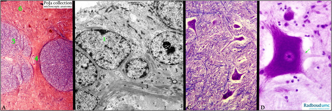
Title: Spinal cord 2
Description:
(A): Silver-stained spinal cord, bovine, with (4) the central canal covered with ependymal cells.
(5) Anterior funiculus. (6) Anterior horn with some large motoneurons.
(B): Electron microscopy, rabbit. Ependymal cells (1) around the central canal which are neighbored by nerve
cells (2) and their axons with conspicuous bundles of longitudinal and cross sectioned neurofilaments (3).
The ependymal cells possess numerous densely stained mitochondria and some short microvilli.
(C): Stain Kluver-Barrera staining, human. Anterior column (ventral grey column) with numerous motoneurons and
their nerve fibers.
(D): Stain cresyl violet, bovine. Anterior column (ventral gray column) with large motoneuron.
Arrow indicates the axon hillock.
Keywords/Mesh: nervous tissue, spinal cord, central canal, gray matter, white matter, motoneuron, ependym, axon, neurofilament, histology, electron microscopy, POJA collection