
11.3 POJA-L3122+3123+3126.
Spinal cord with gray and white matter
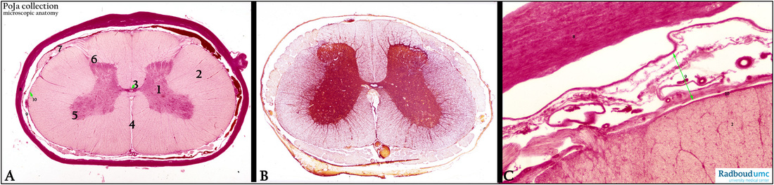
11.3 POJA-L3122+3123+3126
Title: Spinal cord with gray and white matter
Description:
(A, C): Modified silver stain, cervical, bovine.
(A, 1) Gray matter.
(A, 2) White matter.
(3) Central canal located in the commisura grisea.
(4) Anterior media fissure.
(5) Anterior (ventral) gray horn.
(6) Posterior (dorsal) gray horn.
(7) Dorsal root fibers.
The spinal cord is enclosed by the dura (A, 8), arachnoid (A, 9) and pia mater (A, 10), separated from each other by the subdural and subarachnoid spaces. The subarachnoid space is filled with cerebrospinal fluid (CSF).
- The dura mater consists of dense connective tissue.
- The arachnoid is a thin transparent connective tissue layer (spider web appearance) that is lined with an endotheloid cover at the dura side.
- The pia mater is a thin loosely connective tissue layer that is closely situated to the glial limiting membrane and conducts the small capillaries into the nervous tissue.
(B): Silver-stained, midlumbar, bovine. The relative size of the H-figure varies at the various cross-section levels
of the spinal cord. The cross section through the mid-lumbar part of the spinal cord shows identical layers and structure, however, with different sizes. Lumbar segment have massive anterior and posterior horns and contain less white matter than the cervical segments.
(C): Detail of the “meninges” or enclosing layers around the spinal cord. (C, 8) Dura mater. (C, 9) Arachnoid.
Note the numerous blood vessels. (C, 10) Pia mater. (C, 2) White matter showing cross-sectioned myelinated
nerve fibers.
Keywords/Mesh: nervous tissue, spinal cord, spinal cortex, spinal medulla, meninx, dura mater, arachnoidea, pia mater, histology, POJA collection