
12.2.3 POJA-L3381+3382+
2604+3383+2603
Epithelial lining of the eustachian tube in the middle ear
12.2.3 POJA-L3381+3382+2604+3383+2603
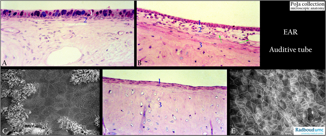
Title: Epithelial lining of the eustachian tube in the middle ear
Description:
(A): Stain alcian blue-PAS, semi-thin plastic section, rabbit.
(B): Stain Haematoxylin-eosin, semi-thin plastic section, near ostium pharyngeum of tube, rabbit.
(C): Scanning electron micrograph, rat
(D): Stain Haematoxylin-eosin, semi-thin plastic section, ostium pharyngeum of tube, rabbit.
(E): Scanning electron micrograph, squamous epithelium of the tube, rat.
- (1) Lining epithelium in varying areas within the middle ear cavity.
- (2) Proper lamina (Lamina propria, layer of connective tissue).
- (B, 3) Outer circumferential lamellae in the osseus part of the tuba. Note proper lamina fused with periost including collagen fibres
and blood vessels (green arrows) followed by the first layers with several bone cells (osteocytes) of the osseus tissue.
In the deeper part the osteocytes are scattered and not yet regularly arranged into so-called osteons or Haversian systems.
- Generally the largest part of the middle ear cavity is composed of flat squamous epithelium (D, E) interspersed with areas of ciliated epithelium (C).
Coursing from different distinct areas ciliated cells are arranged in tracts (A, B) toward the pharyngeal orifice of the eustachian tube (ostium pharyngeum).
Note tall respiratory glandular epithelial cells in (A) and in (B) the decrease in height of the cells tending to layered squamous epithelium close to the tube orifice as depicted in (D).
Keywords/Mesh: middle ear, eustachian tube, auditory tube, respiratory epithelium, ciliated epithelium, histology, electron microscopy,
POJA collection